Artículo original
Vol. 5 Núm. 1 (English)
16 min
Utility of the Pediatric Appendicitis Score (PAS) and C-Reactive Protein (CRP) as diagnostic tools for acute appendicitis in a primary care hospital
Utilidad del Pediatric Appendicitis Score (PAS) y de la Proteína C Reactiva (PCR) como herramientas diagnósticas de apendicitis aguda en el servicio de urgencias de un hospital de primer nivel
Belén Gallego-Calvo1* , Miguel Gallardo-Padilla1, Raúl Castañeda-Vozmediano2, Clara Ramos-Carrillo1, Adoración Clara Granados-Molina1
1Infanta Elena University Hospital, Valdemoro, Madrid, Spain. 2Faculty of Medicine, Francisco de Vitoria University, Madrid, Spain
Correspondencia:
Recepción:
12/11/2025
Aceptación:
14/11/2025
Publicación:
11/03/2026
Métricas
151
Visualizaciones
35
Descargas
Compartir
Compartir
Resumen (ES)
Introducción: El uso de escalas, como el Pediatric Appendicitis Score (PAS), y la medición de la proteína C reactiva (PCR), pueden ser útiles para orientar el grado de sospecha de apendicitis aguda. Este estudio tiene como objetivos comparar la puntuación PAS y PCR entre pacientes con apendicitis aguda y dolor abdominal inespecífico (DAI), analizar un posible punto de corte de riesgo para estas mediciones y evaluar el coste-efectividad de tres modelos de aplicación del PAS.
Pacientes y métodos: Estudio descriptivo retrospectivo en pacientes con sospecha de apendicitis durante 2 años. Se compararon las medianas de PAS y PCR entre grupos, se estableció un punto de corte de riesgo de apendicitis, se analizó el área bajo la curva (AUC) de PAS y PCR individual frente a PAS+PCR combinada, y se realizó un estudio coste-efectividad de 3 modelos de aplicabilidad del PAS según pruebas de imagen.
Resultados: 188 pacientes (127 DAI, 61 apendicitis). Medianas PAS y PCR: 7 [6-8], 2,2 [0,5-4,1] en apendicitis; 4 [3-6], 0,4 [0,1-2] en DAI, (p < 0,001). Punto de corte de riesgo: PAS 6,08 y PCR 0,58 mg/dl. Ningún paciente con PAS ≤ 3 presentó apendicitis. No superioridad del modelo PAS + PCR en la muestra global. PCR ≥ 0,4 mg/dl mayor riesgo de apendicitis en grupo de riesgo intermedio. La estrategia “PAS eficiente” fue la más costoefectiva (ratio costoefectividad 296).
Conclusiones: El PAS es eficaz para estratificar el riesgo de apendicitis. La PCR podría ser de utilidad en el grupo de riesgo intermedio. La implementación del “PAS eficiente” demostró ser una estrategia útil y costoefectiva.
Palabras clave (ES):
Resumen (EN)
Introduction: The use of scales, such as the Pediatric Appendicitis Score (PAS), and the measurement of C-reactive protein (CRP), may help guide the suspicion of acute appendicitis. This study aims to compare PAS and CRP scores between patients with acute appendicitis and those with non-specific abdominal pain (NSAP), analyze potential risk thresholds for these measurements, and assess the cost-effectiveness of three PAS application models.
Patients and methods: A retrospective descriptive study was conducted over two years in patients suspected of appendicitis. The medians of PAS and CRP were compared between groups, a risk threshold for appendicitis was established, the area under the curve (AUC) for individual PAS and CRP models vs combined PAS+CRP was analyzed, and a cost-effectiveness study of three PAS application models based on imaging tests was performed.
Results: A total of 188 patients were included (127 with NSAP, 61 with appendicitis). PAS and CRP medians: 7[6-8], 2.2[0.5-4.1] for appendicitis; 4[3-6], 0.4[0.1-2] for NSAP (p<0.001). Risk thresholds: PAS 6.08, CRP 0.58 mg/dL. No patient with PAS ≤3 had appendicitis. No superiority of the PAS+CRP model in the overall sample. CRP ≥0.4 mg/dL indicated higher risk of appendicitis in the intermediate-risk group. The “efficient PAS” strategy was the most cost-effective (cost-effectiveness ratio 296).
Conclusions: PAS is effective for stratifying appendicitis risk, while CRP may be useful in the intermediate-risk group. The “efficient PAS” implementation proved to be a valuable and cost-effective strategy.
Palabras clave (EN):
Introduction
Right lower quadrant (RLQ) pain is a frequent reason for consultation in pediatric emergency departments, which requires a rigorous differential diagnosis with acute appendicitis(1,2,3,4). Its diagnosis can sometimes be complicated, exposing the patient to complementary tests, usually blood analysis or abdominal ultrasound(3,5,6). In doubtful cases, the patient may undergo tests with greater radiation, such as abdominal CT (computed tomography)(7,8,9). The use of diagnostic scales, such as the Pediatric Appendicitis Score (PAS), together with analytical parameters such as C-reactive protein (CRP), may be useful to guide the degree of suspicion of appendicitis and reduce the number of complementary tests(10,11,12,13). This scale assigns 1 point to the variables: anorexia, nausea/vomiting, pain migration, temperature >38ºC, leukocytes >10,000, neutrophils >75%; and 2 points to the variables: RLQ pain and positive Blumberg sign. Its authors consider scores ≤3 as low risk (no imaging test needed); 4-6 as intermediate risk (imaging tests needed); ≥7 as high risk (consider appendectomy without imaging test)(14,15,16,17,18).
The main objective of this study was to compare the PAS score and the CRP value between patients with acute appendicitis and non-specific abdominal pain (NSAP), analyzing the existence of a risk cutoff point for both in the diagnosis of appendicitis. The total sample and the risk subgroups according to the PAS risk classification were analyzed. Secondary objectives were the comparison of the PAS score and CRP in patients with simple (phlegmonous) and complicated (gangrenous or perforated) appendicitis, as well as conducting a cost-effectiveness analysis based on the imaging tests requested in patients with suspected acute appendicitis, according to 3 situations based on the applicability of the PAS scale (1— real situation applied in the hospital; 2— strict application of PAS; 3— partial application of PAS).
Materials and Methods
A descriptive, retrospective, and inferential study was conducted on patients between 5 and 15 years old attended the emergency department for suspected acute appendicitis at a first-level hospital during the 2021-2022 period. Patients who underwent an abdominal ultrasound were selected, and the variables of the PAS scale were collected from their clinical records. Patients <5 years old, those with an incomplete PAS, and patients with previous appendectomy were excluded. The following variables were collected: sex, age, PAS scale score, numerical value of CRP, radiological diagnosis after imaging test (abdominal ultrasound, abdominal CT scan), and pathology result after appendectomy. Patients were classified as having acute appendicitis based on the result of the imaging tests, distinguishing between simple and complicated. The diagnosis of non-specific abdominal pain (NSAP) was established in cases where no disease was detected by imaging tests. The study was approved by the Clinical Research Ethics Committee of the Fundación Jiménez Díaz prior to its realization.
PAS scores and CRP values between the appendicitis and NSAP groups were compared using the chi-square test or Fisher’s exact test for categorical variables and the Kruskal-Wallis test or the Wilcoxon test for quantitative variables. Scores between patients with simple and complicated appendicitis were also compared. Furthermore, Cramer’s V was estimated to measure the strength of association, and the AUC was evaluated to compare the diagnostic utility of PAS and CRP, individually and combined. The statistical software R (version 4.3) was used.
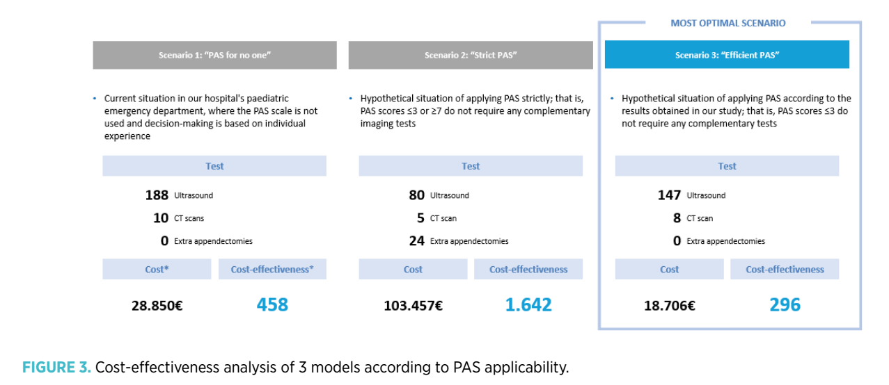
Finally, a cost-effectiveness study was carried out, analyzing the direct cost of imaging tests (abdominal ultrasound: 110 euros; abdominal CT scan: 317 euros), as well as the cost per appendectomy (3,878 euros) in relation to the number of patients diagnosed with appendicitis. The cost-effectiveness analysis ratio (CEAR) was calculated based on 3 possible scenarios according to the applicability of the PAS. In the first, the real situation of our center until then was analyzed; that is, the PAS scale was not used, and clinical decision-making was based on individual experience. The second scenario proposed a hypothetical situation with strict application of the PAS scale (PAS scores ≤3 or ≥7 do not require a complementary imaging test). This second scenario could increase the number of erroneous appendectomies by overtreating patients with a PAS ≥7, so a third hypothetical scenario was proposed, according to which patients with PAS ≤3 were not requested an imaging test, while those with PAS >3 were requested one in all cases.
Results
During the study period, 462 ultrasounds were performed due to suspicion of appendicitis. After applying the inclusion and exclusion criteria, 188 patients (41%) were selected. 106 were male (56.9%), with a mean age of 10.5 years. 125 patients (66.5%) were classified as having non-appendicitis-related diseases (DAI, Diagnóstico de otra patología distinta a Apendicitis) and 63 patients (33.5%) as having acute appendicitis (37 cases of simple appendicitis, 26 cases of complicated appendicitis). There was a 95% correlation between the diagnosis of appendicitis by imaging test and anatomopathological examination.
The medians and interquartile ranges of the obtained PAS score were 7 [6-8] in the appendicitis group and 4 [3-6] in the non-appendicitis-related diseases group (p<0.001, Cramer’s V 0.52), while the CRP values were 2.2 [0.5-4.1] and 0.4 [0.1-2] respectively (p<0.001).
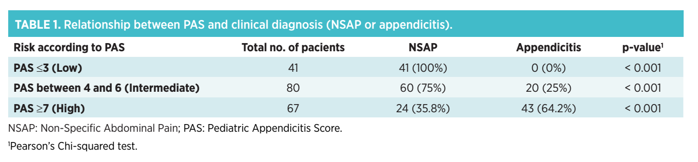
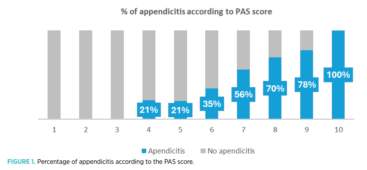
100% of patients with PAS ≤3 (41/41) did not present appendicitis. In the intermediate-risk group, 75% (60/80) of patients with PAS 4-6 were classified as non-appendicitis-related diseases, while, in the high-risk group, 64.2% (43/67) of patients presented appendicitis (Table 1). A higher percentage of appendicitis was observed in relation to PAS scores above 6 (Figure 1). Risk cut-off points of 6.08 for PAS and 0.58 mg/dl for CRP were established.
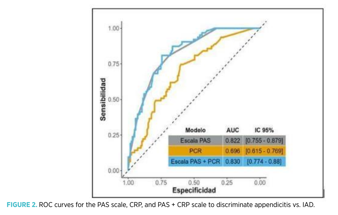
In the comparative analysis using ROC curves between the individual PAS, individual CRP, and combined PAS+CRP models, significant differences in AUC were found between PAS and CRP (p=0.003), but not in the AUC value between individual PAS and PAS+CRP (p=0.853) (Figure 2).
In the sub-analysis of risk groups based on PAS (Table 2), 24.4% (10 out of 41) of patients with PAS ≤3 had CRP values ≥ 0.58 mg/dl, with no case of appendicitis in this group. In the intermediate-risk group, 75% (15/20) of patients with appendicitis had a CRP ≥ 0.58 mg/dl, compared to 40% (24/60) in the non-appendicitis-related diseases group, this association being statistically significant (p=0.007). In the high-risk group, 74.4% (32/43) of patients with appendicitis presented CRP values ≥ 0.58 mg/dl, very similar to those of patients with non-appendicitis-related diseases, where 66.7% (16/24) exceeded this value, this association not being significant (p=0.5).
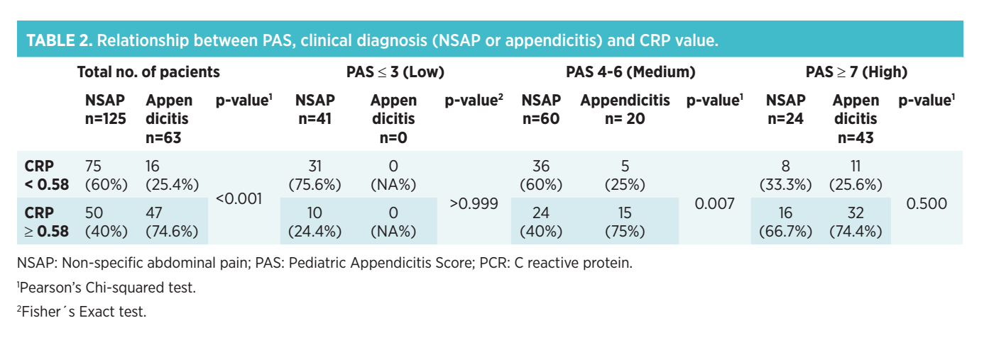
This analysis was performed taking the risk cut-off point for CRP in the general sample. However, when analyzing the optimal CRP cut-off point according to the risk group based on PAS, a CRP value of ≥ 0.4 mg/dl was obtained in the intermediate-risk group and ≥ 2.4 mg/dl in the high-risk group. Considering these values, 90% (18/20) of patients with appendicitis in the intermediate-risk group presented a CRP value ≥ 0.4 mg/dl, compared to 48.3% (29/60) in the non-appendicitis-related diseases group, this association being statistically significant (p=0.001). Furthermore, in the high-risk group, 55.8% (24/43) of appendicitis cases and 37.5% (9/24) of patients with non-appendicitis-related diseases presented a CRP ≥ 2.4 (p=0.151).
Within the group of patients with appendicitis, 58.7% (37/63) were classified as simple. No statistically significant differences were found between simple appendicitis vs. complicated appendicitis in the PAS score (7[6-8] vs. 7.5[7-8], p=0.071) or in the CRP value (2.1 [0.5-3.4] vs. 2.2 [0.7-6.7], p=0.357).
Regarding the results of the cost-effectiveness study, the strategy followed until then in our center presented a cost-effectiveness ratio (CEAR) of 458. Strategy number 2 would imply requesting a lower number of imaging tests (108 ultrasounds and 5 fewer CT scans, relative to the first assumption), but with the performance of 24 extra appendectomies in healthy patients (CEAR 1642). Furthermore, Strategy 3 would imply performing 41 fewer ultrasounds and 2 fewer CT scans compared to Strategy 1, with no incorrectly operated patients and a better cost-effectiveness ratio (CEAR 296). The results of the cost-effectiveness study can be seen in Figure 3.
Discussion
In emergency departments, the diagnostic priority for a child with acute abdominal pain is to rule out acute abdomen, with appendicitis being a common and significant cause of morbidity and mortality in the pediatric population(19,20). Diagnosis in the pediatric population is particularly challenging(3,4,5,6,21).
Nonspecific Abdominal Pain (NAP) is the most common diagnosis at discharge in these patients, with a low rate of organic disease (less than 5%). However, due to the clinical similarity between NAP and appendicitis, additional imaging tests are frequently used, which can lead to misdiagnoses and increased healthcare costs, as well as unnecessary radiation exposure in some cases(5,6,7,8,9).
The use of validated scales such as the Pediatric Appendicitis Score (PAS), in conjunction with other laboratory parameters such as C-Reactive Protein (CRP), can be valuable in clinical decision-making, minimizing iatrogenesis and the costs of imaging tests(10,11,12,13). Goldman(16) classified the PAS risk into three groups, considering scores ≤2 as very low risk and ≥7 as high risk. Subsequent studies indicated that a low-risk PAS (≤3 points) has an appendicitis risk of 2%, while a PAS ≥7 was associated with a risk of 78% to 96%(14,23).
In our sample, the stratification of appendicitis risk according to the PAS showed interesting results. 100% of our patients with low risk (≤3 points) did not present with appendicitis. This finding is particularly relevant in hospitals without a 24-hour on-site radiologist, where the clinical suspicion of appendicitis can lead to the request for imaging tests with higher radiation, such as abdominal CT, or prolong the stay in the emergency department. It is essential to instruct parents about the need to reconsult the emergency department if symptoms worsen and to explain warning signs(22,24).
In the intermediate-risk PAS group, the percentage of appendicitis was 25%. An increase in the number of appendicitis diagnoses was observed with PAS scores ≥7, with higher percentages corresponding to higher PAS scores (56% with PAS = 7; 70% with PAS = 8; 78% with PAS = 9; 100% with PAS = 10)(14). However, the fact that 35.8% (24/67) of high-risk patients did not present with appendicitis necessitates the performance of a confirmatory abdominal ultrasound in this risk group.
Previous studies have analyzed the utility of CRP as a predictive tool in the diagnosis of acute appendicitis. CRP is considered an acute-phase inflammatory reactant that elevates in the first hours of the inflammatory process (4-6 hours, peaking at 12 hours), requires a small sample volume, and is easily accessible in the emergency department(10,25,26,27).
In our study, when evaluating the utility of CRP as a complementary tool to the PAS, we observed that the combined use of PAS+CRP did not prove to be a better predictive model for appendicitis than the isolated use of the PAS scale in the general sample. However, the PAS+CRP application could be especially useful for discriminating a higher risk in the intermediate PAS group. In this group, where diagnostic uncertainties are usually greater, significantly higher percentages of appendicitis than DAI were observed when comparing a CRP value ≥ 0.537 mg/dl (75% versus 40%). When correcting the CRP cut-off point according to PAS risk groups, taking a CRP ≥ 0.4 mg/dl as risk, this difference is even more notable, with percentages of 90% and 48.3% respectively between appendicitis and DAI.
The CRP value was not evaluable in the low-risk PAS group, as there were no patients with appendicitis. It was also less useful in the high-risk PAS group, where, despite presenting a higher percentage in the appendicitis group above the cutoff point, both in the global sample (CRP ≥ 0.537 mg/dl) and in the subgroup analysis of the PAS (CRP ≥ 2.4 mg/dl), the values approached those observed in the DAI group with statistically non-significant differences.
Based on the obtained results, a cost-effectiveness study on the application of the PAS scale was proposed based on 3 models. In our emergency department of a level 1 hospital, no appendicitis prediction scales were used at the time of the study to stratify the risk of patients with abdominal pain.
In the comparative analysis, strategy number 2 was not found to be more beneficial than the initial strategy (strategy 1). Although the strict application of the PAS scale significantly reduced the number of imaging tests requested, the high number of healthy patients with an erroneous diagnosis of appendicitis (32% in the high-risk group) proved not to be a viable option. However, all patients with PAS ≤ 3 were correctly diagnosed as healthy, which is consistent with similar studies reporting appendicitis percentages of <2%(23) in this group. This suggests that strategy 3, which we call “efficient PAS,” is very promising, as it allows for resource savings in the low-risk group without misdiagnoses and improves the cost-effectiveness ratio.
Limitations include its retrospective nature and the assignment of appendicitis or DAI cases based on ultrasound diagnosis. However, the high correlation (95%) between ultrasound and anatomopathology minimizes this bias. More prospective studies are needed to confirm our hypotheses.
Conclusion
The use of the PAS scale is a useful tool that allows for better stratification of patients at risk of appendicitis. In our sample, no patient with a PAS ≤3 presented with appendicitis, and only 25% of the intermediate-risk patients did. In contrast, higher scores in the risk group were associated with a greater risk of presenting with appendicitis. The incorporation of CRP did not prove to be more useful compared to the isolated use of the PAS scale in the general sample, but it did allow us to better guide intermediate-risk patients, where a CRP cut-off point ≥ 0.4 mg/dl was significantly associated with a higher risk of appendicitis. We consider that the application of the “efficient PAS” can optimize clinical decision-making, minimizing the average length of hospital stay for low-risk patients and significantly saving healthcare resources.
Funding
No funding was received for the research, authorship, and/or publication of this article.
Authors’ contributions
Belén Gallego Calvo: Formal analysis, Writing – review & editing, Writing – original draft, Data curation, Methodology, Visualization
Miguel Gallardo Padilla: Writing – review & editing, Writing – original draft, Validation
Raúl Castañeda Vozmediano: Methodology, Formal analysis
Clara Ramos Carrillo: Writing – original draft, Data curation, Writing – review & editing
Adoración Clara Granados Molina: Writing – review & editing, Validation, Writing – original draft, Supervision, Data curation
Conflicts of interest
The authors declare that there are no commercial or financial conflicts of interest for this research.
Ethical declarations
This work (original article, clinical trial, research with human/animal subjects, etc.) complies with research regulations and has the approval of an Ethics Committee. The authors declare that they have the corresponding official document or approval code.
Use of artificial intelligence tools
I declare that no artificial intelligence tool (such as ChatGPT, Copilot, Gemini, or others) has been used in the writing, analysis, or review of this article.
References
- Alonso Cadenas JA, de la Torre Espí M. Diagnóstico y tratamiento del dolor abdominal agudo (abdomen agudo) en Urgencias. Protoc diagn ter pediatr. 2020;1:197-213.
- Astobiza E. Dolor abdominal. En: Benito J, Mintegi S, Sánchez J, editores. Diagnóstico y tratamiento de urgencias pediátricas. 5ª ed. Madrid: Panamericana; 2016:210-20.
- Lipsett SC, Bachur RG. Current Approach to the Diagnosis and Emergency Department Management of Appendicitis in Children. Pediatr Emerg Care. 2017;33:198-205.
- Peña Quintana L, Beltrà Picó R. Dolor abdominal agudo. En: Cruz. Tratado de Pediatría, 11ª ed. Madrid: Panamericana; 2014:1465-8.
- Rassi R, Muse F, Cuestas E. Apendicitis aguda en niños menores de 4 años: un dilema diagnóstico [Acute appendicitis in children under 4 years: a diagnostic dilemma]. Rev Fac Cien Med Univ Nac Córdoba. 2019;76(3):180-4. Available from: https://doi.org/10.31053/1853.0605.v76.n3.23661.
- Rothrock SG, Pagane J. Acute appendicitis in children: emergency department diagnosis and management. Ann Emerg Med. 2000;36(1):39-51. Available from: https://doi.org/10.1067/mem.2000.105658.
- Callahan M, Rodríguez D, Taylor G. CT of appendicitis in children. Radiology. 2002;224:325-32. Available from: https://doi.org/10.1148/radiol.2242010998.
- Rodríguez-Cuellar E, Gutiérrez Andreu M, Gómez Rodríguez P, Alcalde Escribano J, Cruz Vigo F. Impacto de los métodos de diagnóstico por imagen en la apendicectomía de urgencia. Rev Calid Asist. 2010;25(5):308-11. Available from: https://doi.org/10.1016/j.cali.2009.12.007.
- Weston AR, Jackson TJ, Blamey S. Diagnosis of appendicitis in adults by ultrasonography or computed tomography: a systematic review and meta-analysis. Int J Tech Asses Health Care. 2005;21:368-79.
- Prada Arias M, Salgado Barreira A, Montero Sánchez M, Fernández Eire P, García Saavedra S, Gómez Veiras J, et al. Appendicitis versus non-specific acute abdominal pain: Paediatric Appendicitis Score evaluation. An Pediatr (Barc). 2018;88(1):32-8. Available from: https://doi.org/10.1016/j.anpedi.2017.01.006.
- Kulik DM, Uleryk EM, Maquire JL. Does this child have appendicitis? A systematic review of clinical prediction rules for children with acute abdominal pain. J Clin Epidemiol. 2013;66(1):95-104.
- Pogorelić Z, Rak S, Mrklić I, Jurić I. Prospective validation of Alvarado Score and Pediatric Appendicitis Score for the diagnosis of acute appendicitis in children. Pediatr Emerg Care. 2015;31(3):164-8. Available from: https://doi.org/10.1097/PEC.0000000000000375.
- Juanes de Toledo B, Martínez Rubio MV. Escalas de predicción clínica en apendicitis aguda: ¿son útiles? Evid Ped. 2015;11:45.
- Bhatt M, Joseph L, Ducharme FM, Poonai N, Tassone E, Gore G, et al. Prospective validation of the pediatric appendicitis score in a Canadian pediatric emergency department. Acad Emerg Med. 2009;16(7):591-6.
- Samuel M. Pediatric Appendicitis Score. J Pediatr Surg. 2002;37(6):877-81.
- Goldman RD, Carter S, Stephens D, Gorelick M. Prospective validation of the pediatric appendicitis score. J Pediatr. 2009;154(2):308-9.
- Hatcher-Ross K. Sensitivity and specificity of the Pediatric Appendicitis Score. J Pediatr. 2009;154(2):308.
- Planella Cornudella M, Pociello Almiñana N, Domingo Ruiz A, Bringue Espuny X, Rue Monne M, Sole Mir E. Utilidad del Pediatric Appendicitis Score y la ecografía abdominal en el proceso diagnóstico de apendicitis aguda [Utility of Pediatric Appendicitis Score and abdominal ultrasound in the diagnostic process of acute appendicitis]. Cir Pediatr. 2019;32(3):128-34.
- Barker PA, Jutley RS, Youngson GG. Hospital re-admission in children with non-specific abdominal pain. Pediatr Surg Int. 2002;18(5-6):341-3.
- Alvarez Bernaldo De Quirós M, Sanz Villa N, Cortés Gómez MJ. Prospective study of 288 cases of acute appendicitis during childhood: characteristics in preschool children. An Esp Pediatr. 1997;46:369-72.
- Hao TK, Chung NT, Huy HQ, Linh NT, Xuan NT. Combining ultrasound with a pediatric appendicitis score to distinguish complicated from uncomplicated appendicitis in a pediatric population. Acta Inform Med. 2020;28(2):114-8. Available from: https://doi.org/10.5455/aim.2020.28.114-118.
- Pennel DJ, Goergen N, Driver CP. Nonspecific abdominal pain is a safe diagnosis. J Pediatr Surg. 2014;49(11):1602-4. Available from: https://doi.org/10.1016/j.jpedsurg.2014.06.014.
- Brandt M. Acute appendicitis in children: clinical manifestations and diagnosis. In: UpToDate. Wiley J, editor. UpToDate; 2023. Available from: URL.
- Velasco Zúñiga R, Luis J, Arribas F, Montes SP, Natali M, Fernandez C, et al. Application of Pediatric Appendicitis Score on the Emergency Department of a Secondary Level Hospital. Available from: http://www.pec-online.com.
- Sondenaa K, Buan B, Soreide JA, Nysted A, Andersen E, Nesvik I, et al. Rapid c-reactive protein (CRP) measurements in the diagnosis of acute appendicitis. Scand J Clin Lab Invest. 1992;52(7):585-9. Available from: https://doi.org/10.1080/00365519209115500.
- Gurleyik E, Gurleyik G, Unalmiser S. Accuracy of serum C-reactive protein measurements in diagnosis of acute appendicitis compared with surgeon’s clinical impression. Dis Colon Rectum. 1995;38:1270-4.
- Stefanutti G, Ghirardo V, Gamba P. Inflammatory markers for acute appendicitis in children: are they helpful? J Pediatr Surg. 2007;42(5):773-6. Available from: https://doi.org/10.1016/j.jpedsurg.2006.12.028.
Explorar
Artículos
Volúmenes
Sobre la revista
Más leídos
Emergencias Pediátricas
Emergencias Pediátricas
Emergencias Pediátricas
1º Congreso Mundial de Emergencias Pediátricas
Emergencias Pediátricas
Una nueva mirada a la vía aérea pediátrica: secuencia de intubación diferida como alternativa segura en situaciones de urgencias
Emergencias Pediátricas