Artículo original
Vol. 5 Núm. 2 (English)
15 min
Traumatic brain injury in the pediatric emergency room: less radiation, more observation
Traumatismo craneoencefálico en urgencias pediátricas: menos radiación, más observación
Valentina Ruiz-Blanco1*, Denisse-Alejandra Moggia-Morla1, Sonia Cañadas-Palazón2
1Department of Pediatrics, Vall d'Hebron University Hospital, Barcelona, Spain. 2Pediatric Emergency Unit, Vall d'Hebron University Hospital, Barcelona, Spain
Correspondencia:
Recepción:
26/10/2025
Aceptación:
21/01/2026
Publicación:
26/06/2026
Métricas
102
Visualizaciones
3
Descargas
Compartir
Compartir
Resumen (EN)
Introduction: Traumatic brain injury (TBI-TCE) is a common pathology in pediatrics. It is important to stratify the risk of developing an intracranial injury (ICI-LIC) after a TBI and select the high-risk patients for additional diagnostic tests.
Objective: To compare the risk stratification of ICI and the management of patients with TBI in the emergency department in 2022, based on two protocols (1-2017; 2-2023), evaluating the impact on the number of diagnostic imaging performed and the injuries detected.
Methods: A retrospective study was conducted on patients aged 0-18 years diagnosed with TBI in a 3rd-level pediatric emergency department during 2022. A descriptive analysis of epidemiological and clinical data, risk classification, and management was performed based on protocol 1-2017. A theoretical comparison was then made by applying Protocol 2-2023 to the same patients to assess changes in classification and management.
Results: A total of 811 patients were evaluated. 633 (78%) were classified as low risk for IBI, 109 (13.5%) as intermediate risk, and 69 (8.5%) as high risk. Nineteen skull X-rays (2.2%) and 87 head CT scans (10.7%) were performed, detecting 19 skull fractures, 13 ICIs, and 9 extracranial bleedings. Applying protocol 2-2023 would have resulted in no X-rays and a reduction of CTs to 72 (8.8%), with all ICIs still being diagnosed.
Conclusions: The updated protocol maintains diagnostic effectiveness in identifying ICIs while reducing the number of imaging studies, thereby minimizing their potential adverse effects.
Palabras clave (EN):
Resumen (ES)
Introducción: El traumatismo craneoencefálico (TCE) es una patología frecuente en pediatría. Es importante estratificar el riesgo de presentar una lesión intracraneal (LIC) tras un TCE y seleccionar a los pacientes de mayor riesgo para realizar exploraciones complementarias.
Objetivo: Comparar la estratificación de riesgo de LIC y el manejo de los TCE en urgencias en el 2022 según 2 protocolos (1-2017; 2-2023), evaluando el impacto en el número de exploraciones complementarias realizadas y las lesiones detectadas.
Métodos: Estudio retrospectivo de los pacientes de 0 a 18 años diagnosticados de TCE en urgencias pediátricas en un hospital terciario durante el 2022. Se realizó un análisis descriptivo de datos epidemiológicos y clínicos, la clasificación de riesgo y el manejo realizado según el protocolo 1-2017. Posteriormente, se asoció una comparación teórica de la clasificación y el manejo de dichos pacientes tras aplicar el protocolo 2-2023.
Resultados: De 811 pacientes, 633 fueron clasificados como riesgo bajo de LIC (78%), 109 como intermedio (13,5%) y 69 como alto (8,5%). Se realizaron 18 radiografías de cráneo (2,2%) y 87 tomografías (TAC) de cráneo (10,7%), diagnosticando 19 fracturas de cráneo, 13 lesiones intracraneales y 9 extracraneales. Aplicando el protocolo 2-2023, no se habrían realizado radiografías y hubiera disminuido el número de TACs a 72 (8,8%), p <0,001; todas las LIC hubieran sido diagnosticadas (p 0,67).
Conclusiones: La actualización del protocolo mantiene una eficacia diagnóstica respecto al hallazgo de LICs y a su vez reduce el número de exploraciones complementarias en los TCE, disminuyendo así sus efectos adversos.
Palabras clave (ES):
Introduction
Traumatic brain injury (TBI) is a very frequent reason for consultation in pediatric emergency departments; it represents 3-6% of visits and 1-2% of admissions, with a higher prevalence in children under 2 years of age.
Most pediatric TBIs are mild and do not involve intracranial lesions (ICLs); fewer than 10% present them, and fewer than 1% require urgent neurosurgical intervention(1-2). Clinically significant intracranial lesion (csICL) is defined as one that meets any of the following characteristics: requiring neurosurgical intervention, requiring intubation >24h, requiring hospital admission >48h, or causing the death of the patient(3). A careful evaluation of TBIs is essential, as some can lead to high mortality and morbidity, with the appearance of long-term sequelae. Mortality is higher the younger the patient’s age, reaching up to 30% in severe TBIs(4).
The imaging of choice when an ICL is suspected is a computed tomography (CT) scan of the head; however, this test carries significant associated risks, mainly from ionizing radiation. This is associated with an increased risk of long-term carcinogenesis in children, given the high sensitivity of their developing organs and a longer remaining life expectancy(6). Another important risk can be the need for sedation, especially in infants and younger children(1). It is emphasized that up to 80-90% of CT scans in mild trauma are normal or may show non-clinically relevant lesions (e.g., soft tissue hematomas).
Skull radiography (skull X-ray) has fallen into disuse given its poor diagnostic performance for detecting ICLs and the possibility of requiring further study with a CT scan in cases of pathological findings or diagnostic doubts(7).
It is important to have updated protocols that allow the pediatrician to identify risk cases to act early and, at the same time, minimize the performance of imaging tests in patients with a lower risk.
In 2023, a review of TBI assessment scales and subsequent studies on them was conducted in the pediatric department. Large-scale studies were used as a basis, such as the Children’s Head Injury Algorithm for the Prediction of important Clinical Events (CHALICE) in 2006(9), the Pediatric Emergency Care Applied Research Network (PECARN) in 2009(3), and the Canadian Assessment of Tomography for Childhood Head Injury (CATCH) in 2010(10), which were performed to validate prediction rules for classifying TBIs according to the risk of presenting csICL, thus attempting to reduce unnecessary head CT scans. The primary objective is to find variables with high sensitivity and high negative predictive value (near 100% in all three studies), prioritizing a correct medical history and a prolonged period of clinical observation over performing a head CT scan if these variables are met(11). Following this review, the in-hospital TBI protocol at H. Vall d’Hebron was updated.
The objective of this study is to compare the classification and management of TBIs in the emergency department under the previous protocol with the theoretical classification and management these same patients would have had using the updated protocol, evaluating the impact on the number of complementary examinations performed and the lesions detected.
Material and Methods
A retrospective study was conducted of patients aged 0-18 years diagnosed with TBI in the pediatric emergency department during the year 2022 through a review of medical records. The study was approved by the hospital’s clinical research ethics committee.
Exclusion criteria included relevant history that could alter the management proposed by the protocol, such as coagulopathies, neurosurgical history, or neurological diseases requiring individualized management, as well as patients with suspicion of non-accidental trauma.
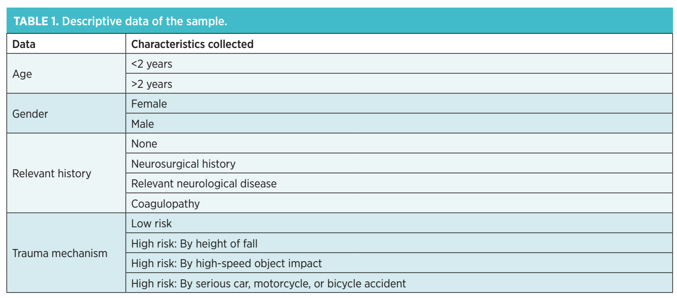
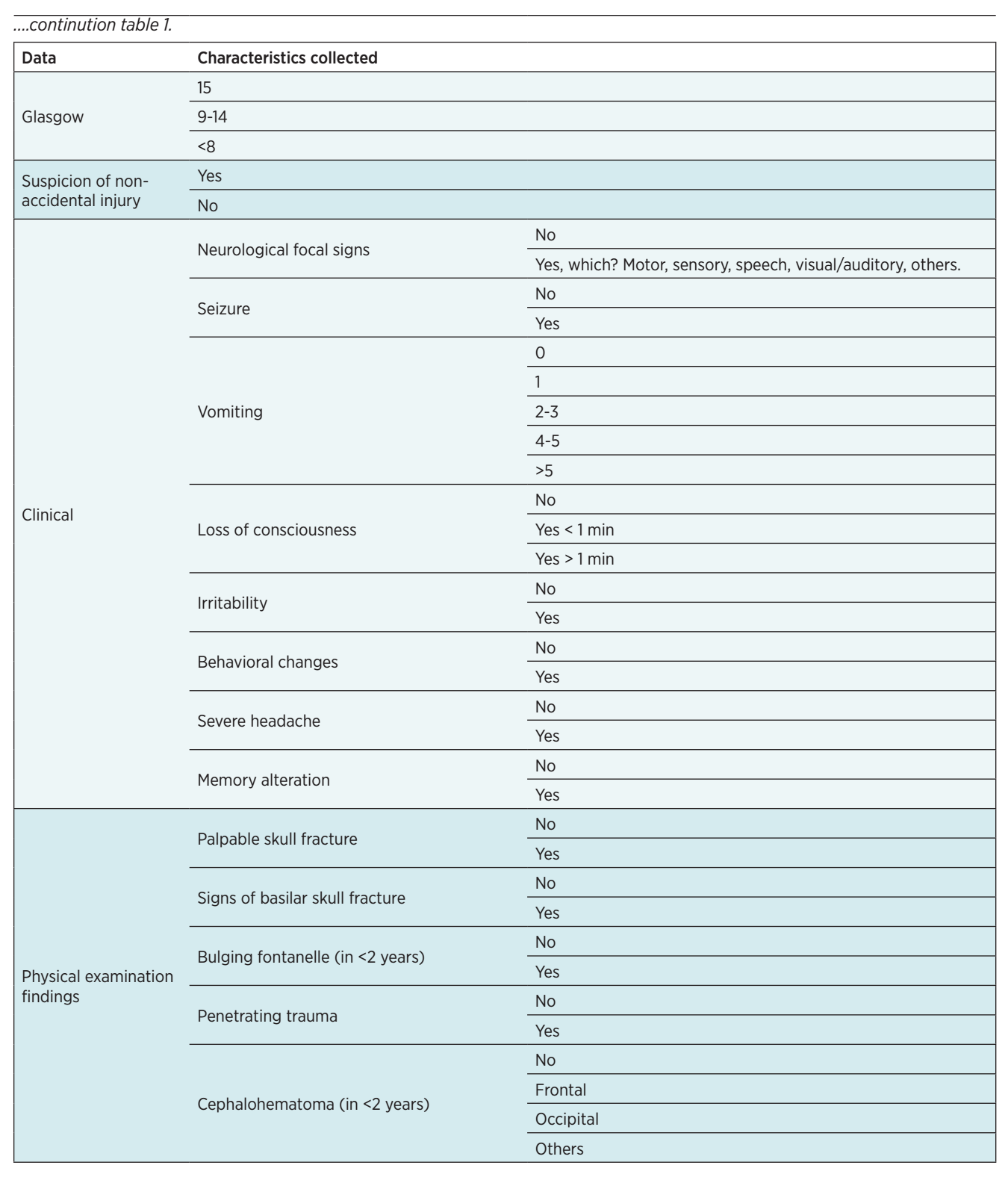
The epidemiological characteristics of the patients, the TBI risk classification assigned according to the trauma’s mechanism of injury, post-traumatic symptoms and clinical findings, as well as the management performed and imaging findings, were analyzed. During this year, the hospital’s TBI management protocol, last revised in 2017, was in effect. The data collected from each medical record are summarized in Table 1.
Subsequently, a comparison was made between the theoretical classification and proposed management if the same patients had been treated under the protocol updated in 2023. This comparison is hypothetical and cannot replicate clinical decision-making or real-world patient evolution conditions.
For both protocols, the initial management according to risk classification is as follows:
- High risk: Head CT scan.
- Intermediate risk: In-hospital observation (4 to 6 hours). A head CT scan is considered if more than one intermediate risk criterion is present, or in case of persistent symptoms or worsening during the observation period.
- Low risk: Discharge home with recommendations and instructions for return consultation.
The main differences between the risk classification of both protocols are:
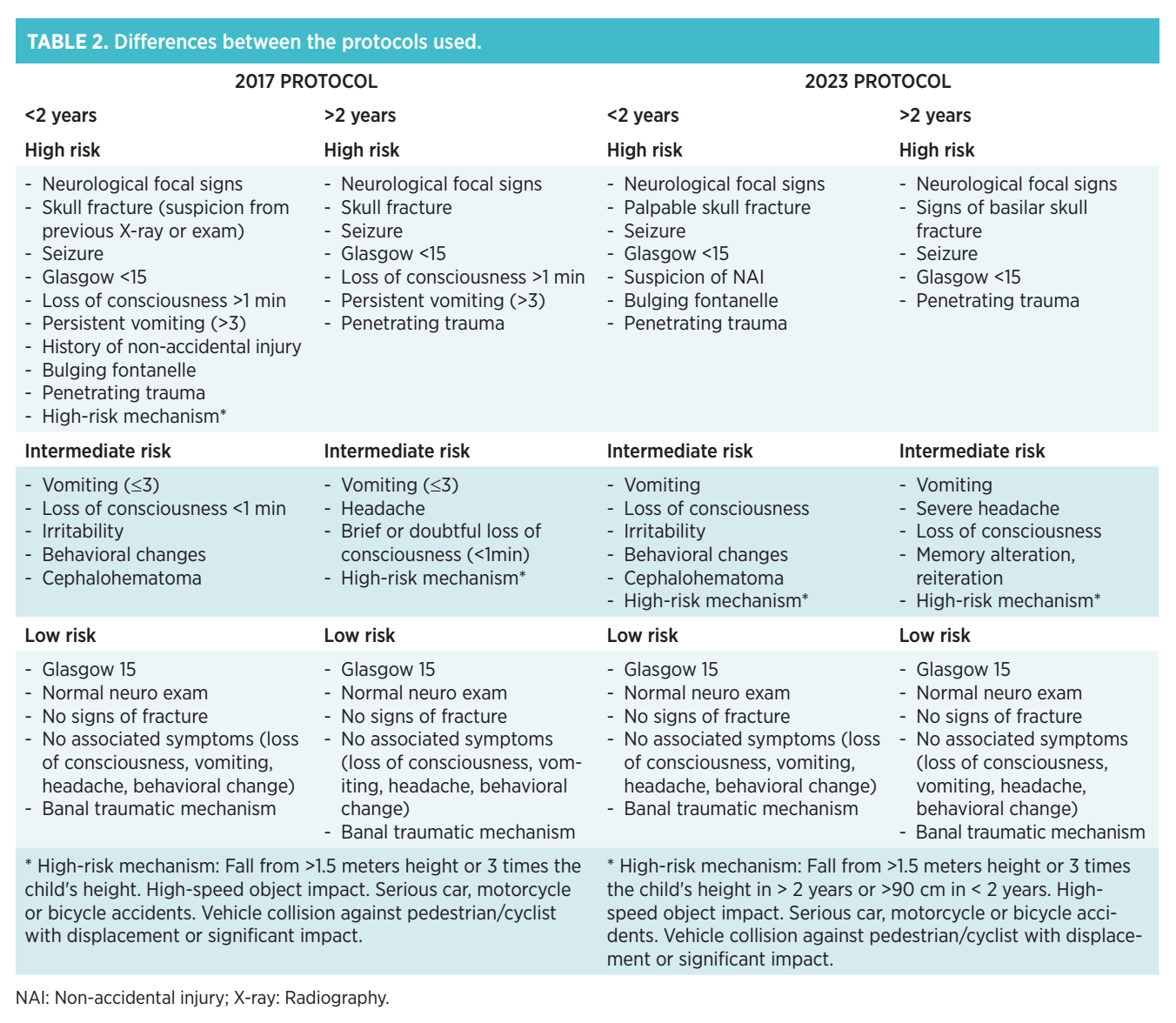
- The high-risk mechanism includes a fall from more than 1.5 meters in >2 years or more than 90 cm in <2 years or 3 times the child’s height; an impact with a high-speed object or serious car, motorcycle, or bicycle accidents; and collisions against a pedestrian or cyclist with displacement or significant impact; previously this was classified as a high-risk criterion for <2 years and intermediate risk for >2 years. In the new protocol, it is considered intermediate risk for both age groups. This is because the presence of a high-risk mechanism alone without other clinical signs implied performing a head CT scan, and it has been shown that in these cases, conducting a regulated in-hospital observation of 4-6 hours significantly reduces complementary examinations without affecting the patient’s prognosis.
- The number of vomits is currently not considered a criterion for greater TBI severity, so it becomes an intermediate risk criterion.
- The criterion of memory alteration or reiteration is included in the intermediate risk, as it has been observed that associating this criterion with other symptoms such as headache, vomiting, and/or a high-risk mechanism increases the probability of presenting a csICL.
These modifications are in accordance with the update of the SEUP TBI protocol published in February 2024(5) and are shown in Table 2.
Results
A total of 851 medical records were reviewed; after applying exclusion criteria, 811 patients were included. They were classified by sex: 513 male (63.3%) and 298 female (36.7%); and by age: 217 under 2 years (26.8%) and 594 aged 2 years or older (73.2%). 91.7% (744) of consultations were for traumas with low-risk injury mechanisms.
According to the 2017 protocol, 633 patients were classified as low risk (78%), 109 as intermediate risk (13.5%), and 69 as high risk (8.5%). 18 skull X-rays and 87 head CT scans were performed. A total of 19 skull fractures were detected, 18 by CT and 1 by skull X-ray, 3 intracranial hematomas, 5 intracranial lesions other than hematomas (subarachnoid bleeding, petrous bone fractures, and one orbital fracture), and 9 extracranial lesions consisting of subgaleal and epicranial bleeding. The characteristics of the patients who presented with lesions are summarized in Table 3.
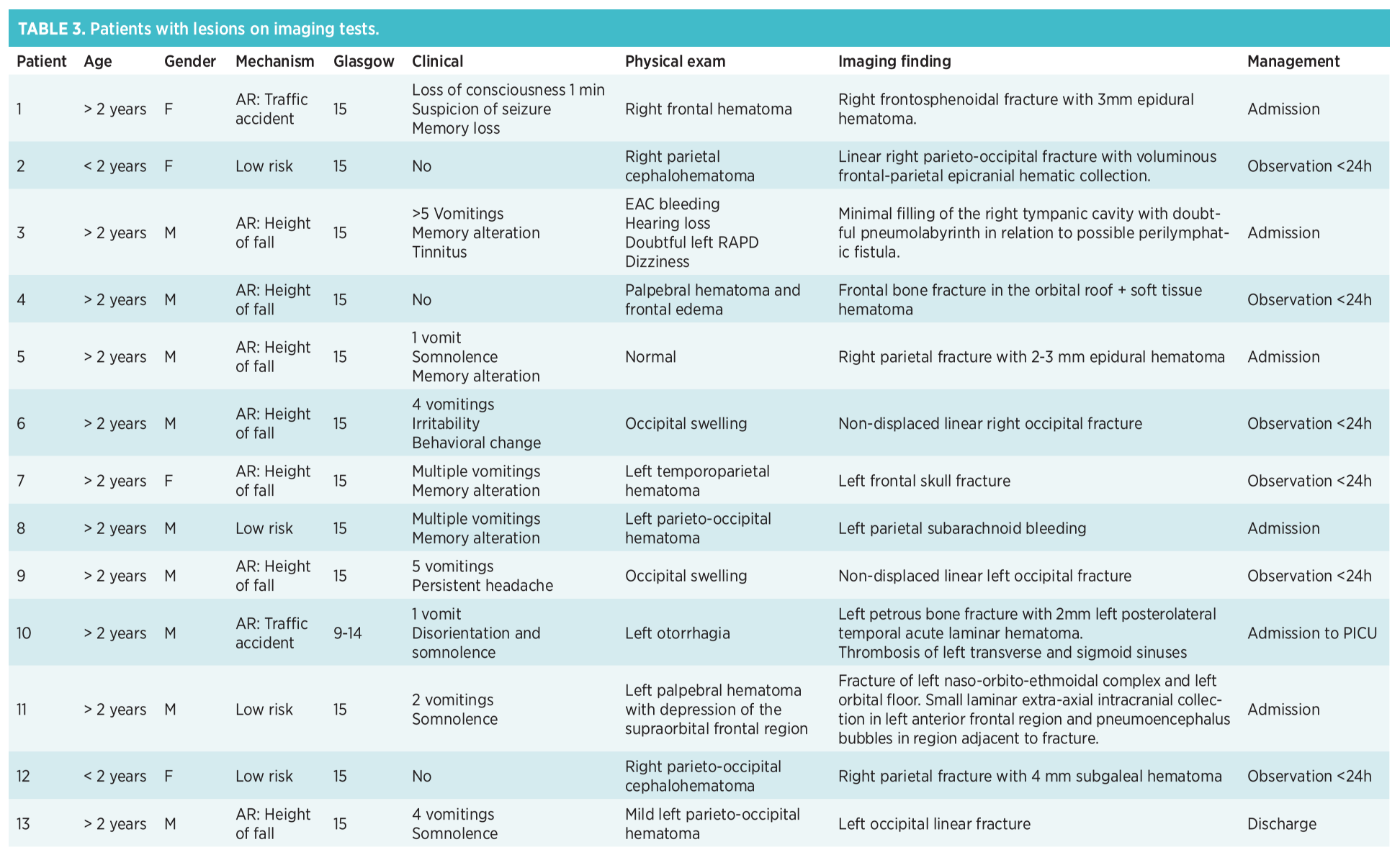
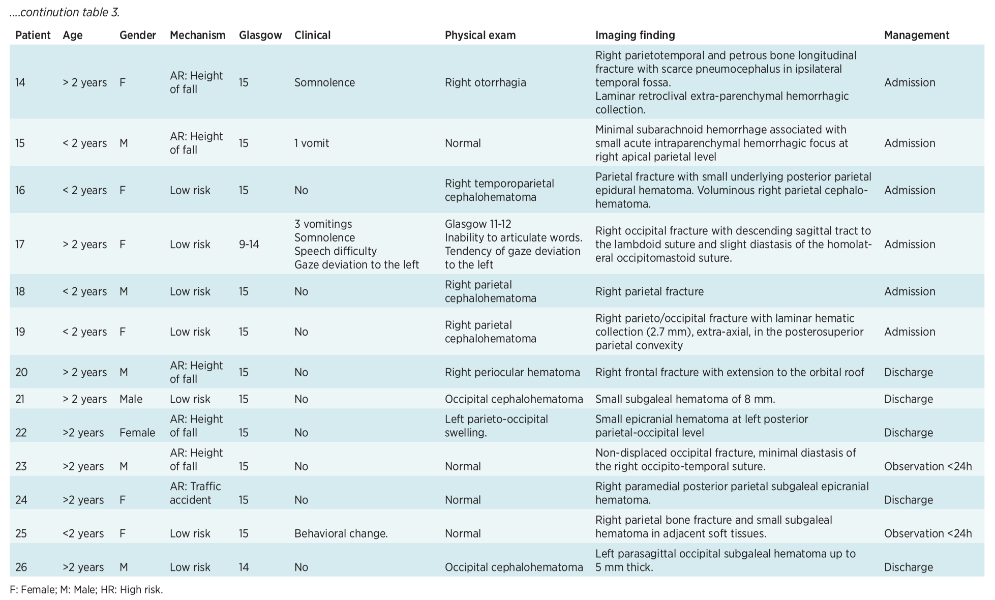
Among patients with ICL findings, 5 presented a high-risk mechanism: 3 due to the height of the fall and 2 due to a traffic accident. The symptoms most frequently associated with ICL were vomiting, somnolence, and memory alteration (amnesia of the episode).
Regarding the physical examination, the most striking feature was the presence of non-frontal cephalohematomas. 3/13 (23%) patients were considered csICL. No statistically significant differences were found in terms of ICL detection between the two protocols (p 0.67).
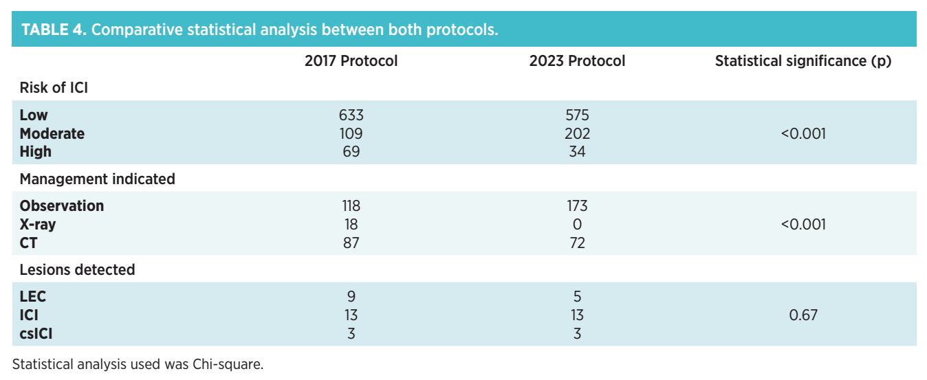
Subsequently, a theoretical comparison of the assigned risk and management of the same 811 patients was performed applying the updated protocol. According to the 2023 protocol, 575 patients would have been classified as low risk (70.8%, previously 78%), 202 as intermediate risk (25%, previously 13.5%), and 34 as high risk (4.2%, previously 8.5%). It was observed that in 39 cases, a lower theoretical risk would be assigned than in real-world management: 37 cases from high to intermediate and 2 from intermediate to low; in 57 cases, a higher risk than previously would be assigned; however, management was not significantly modified.
Applying the new protocol, no skull X-ray would have been performed, and 72 head CT scans would have been performed (15 fewer than with the previous protocol) and a greater number of observations (173 vs. 118), these differences being statistically significant (p <0.001). This would have mainly reduced tomographies reported as absence of ICL; however, 3 subgaleal hematomas would not have been detected, one of them associated with a parietal fracture, to which in-hospital observation management would have been applied. All CT scans with ICL results would have been performed equally. The results are summarized in Table 4.
The 35 patients in whom an imaging test (skull X-ray or CT) would have been omitted were reviewed, and it was observed that none of them subsequently returned for clinical worsening nor required additional tests, taking into account that follow-up could not be performed for one of them due to their location. 25 of them were discharged directly from emergency, 9 were admitted for 12 hours for being under 3 months old, and, finally, only 1 patient was admitted for 24 hours due to a parietal fracture finding with a 2 mm subgaleal hematoma (previously mentioned), without requiring additional management.
Discussion
TBI is a frequent reason for consultation, which requires protocols that help the clinician decide on management in emergency departments, including the decision to perform imaging tests(8).
After classifying a patient as intermediate risk, this is where the most variability in actions is observed, as the decision depends largely on the treating physician’s judgment. It is here where clearer recommendations on which patients benefit most from a CT scan are most relevant(13). It is estimated that approximately 30% of TBIs are categorized as “intermediate risk”; in our study, 25%, and of these, 0.9%, regardless of age, will present a csICL.
One criterion used in our protocol 2-2023 to prioritize imaging over observation is the presence of more than one intermediate risk criterion. Yura Ko et al. in their 2019 study(13) describe that for patients with intermediate-risk TBI, an isolated PECARN variable can imply performing imaging tests in 28.5-71.7% and show clinically relevant results only in 0.2-1.4% (need for neurosurgery in 0-0.5%). However, the combination of 2 variables, one being a high-risk mechanism along with the presence of cephalohematoma, vomiting, and/or loss of consciousness, increases the risk of csICL up to 2.6-3%, which could guide us when requesting images in these groups.
The results of our study allow us to reaffirm that updating management protocols in accordance with the aforementioned studies allows for a reduction in complementary imaging while maintaining diagnostic efficacy and prioritizing hospital observation(12).
Regarding in-hospital observation, the results obtained in our study also coincide with other studies, one of them prospective, conducted in Australia in 2020(14) involving 10 emergency departments, which concluded that a planned observation for intermediate-risk, and sometimes high-risk, TBIs reduce the use of imaging tests (4.4% vs. 13.3%) without significant differences in pathological results (between 0.5-1.1%) or increasing the risk of missing a csICL diagnosis in the observation group. Additionally, it supports the use of clinical scales for TBI classification.
In 2011, another study in the United States(15) divided its cohort into a group that underwent an observation period before the decision to perform complementary tests versus a group in which observation was not chosen for indicating or dismissing the test. A decrease in CT use was described in the first group: 31.1% vs. 35% (difference -3.9% with a 95% CI, in our case a 2% decrease), which implies the elimination of 39 CT scans per 1000 patients presenting to emergency for TBI. They also concluded that the risk of ICL in intermediate-category TBI is 0.9% and the observation period allows assessing if patients show clinical improvement and thus being able to dispense with imaging tests. J. Homme describes in his study(2) that with each hour of observation in these patients, CT performance is reduced by up to 70%. Observation time across studies varies between 2-6 hours, and it is reported that up to 96.5% of intracranial hemorrhages are detected in the first 6 hours.
According to a study published in 2018(16), patients who were observed in the emergency department underwent CT less frequently compared to those who were not (5% versus 34%); likewise, it was described that with each hour of observation of a patient after a TBI, the ratio of requested CTs in intermediate-risk TBIs decreases up to 72%, similar to the previously mentioned study.
The description in the literature coincides with the low percentage of ICL in our sample and as we have evidenced, supports conservative management prioritizing clinical observation whenever lower-risk criteria are met(17).
Conclusion
This study corroborates the importance of an adequate medical history and physical examination to properly classify patients into the corresponding risk, as well as the major role played by active clinical observation in approaching patients with TBI, especially those classified within intermediate risk. Rigorously applying prediction rules allows us to minimize unnecessary imaging tests, without increasing the number of undiagnosed ICLs.
Additionally, we emphasize the importance of having updated protocols and the need to conduct multicenter prospective studies on the topic that reinforce the findings described in order to provide the best and safest care to our patients.
Funding
No funding was received for the research, authorship, and/or publication of this article.
Author contributions
Valentina Ruiz Blanco: Conceptualization, Methodology, Writing - original draft, Writing - review & editing, Visualization.
Denisse Alejandra Moggia Morla: Investigation, Formal analysis, Writing - original draft, Writing - review & editing, Visualization.
Sonia Cañadas Palazón: Supervision, Project administration.
All authors critically reviewed the manuscript, approved the final version, and take responsibility for its content.
Conflicts of interest
The authors declare that there is no commercial or financial conflict of interest for this research.
Ethical approval document
This work (original article, clinical trial, research with humans/animals, etc.) complies with research regulations and has been approved by an Ethics Committee. I possess the official document or corresponding approval code.
Use of artificial intelligence tools
The authors declare that no artificial intelligence tool (such as ChatGPT, Copilot, Gemini, or others) has been used in the writing, analysis, or review of this article.
References
- Rivero R, Curran IL, Hellmann Z, Carroll M, Hornick M, Solomon D, et al. Unnecessary Scans Lead to Unnecessary Re-scans: Evaluating Clinical Management of Low and Intermediate Risk Pediatric Traumatic Brain Injuries. J pediatr surg, 2025; 60(2): 162097.
- Homme J. Pediatric Minor Head Injury 2.0: Moving from Injury Exclusion to Risk Stratification. Emerg Med Clin N Am, 2018; 36(2): 287–304.
- Kuppermann N, Holmes J, Dayan PS, Hoyle JD, Atabaki SM, Holubkov R, et al. Pediatric Emergency Care Applied Research Network (PECARN). Identification of children at very low risk of clinically-important brain injuries after head trauma: a prospective cohort study. Lancet. 2009; 374(9696): 1160–1170.
- Fernández F, González E. Traumatismo craneoencefálico. Pediatr Integral. 2024; 28(1): 7 – 16.
- Sociedad Española de Urgencias de Pediatría (SEUP). Protocolos diagnósticos y terapéuticos en urgencias de pediatría. 18. Traumatismo craneal. 4ª Edición, 2024. Disponible en: https://seup.org/wp-content/uploads/2024/04/18_Trauma_craneal_4ed.pdf
- Scott J, Grewal T, Brewster S, Khan A. Optimizing imaging in the pediatric trauma patient, part 1: head and neck trauma. Pediatr Emerg Med Pract. 2021; 18(Suppl 8): 1-39.
- Rello-Saltor V, Garcés-Montolio L, Gander R, Cañadas-Palazón S, Velasco-Puyó P. Maneig del traumatisme cranioencefàlic a urgències. Són necessàries les proves d’imatge?. Pediatr Catalana. 2018; 78(1): 15-24.
- Hess EP, Homme JL, Kharbanda AB, Tzimenatos L, Louie JP, Cohen DM, et al. Effect of the Head Computed Tomography Choice Decision Aid in Parents of Children With Minor Head Trauma: A Cluster Randomized Trial. JAMA netw open. 2018; 1(5): e182430.
- Dunning J, Daly JP, Lomas JP, Lecky F, Batchelor J, Mackway- Jones K & Children’s head injury algorithm for the prediction of important clinical events study group. Derivation of the children’s head injury algorithm for the prediction of important clinical events decision rule for head injury in children. Arch Dis Child. 2006; 91(11): 885–891.
- Osmond MH, Klassen TP, Wells GA., Correll R, Jarvis A, Joubert G, et al. Pediatric Emergency Research Canada (PERC) Head Injury Study Group. CATCH: a clinical decision rule for the use of computed tomography in children with minor head injury. CMAJ: Can Med Assoc J. 2010; 182(4): 341–348.
- Babl FE, Borland ML, Phillips N, Kochar A, Dalton S, McCaskill M, et al. Paediatric Research in Emergency Departments International Collaborative (PREDICT). Accuracy of PECARN, CATCH, and CHALICE head injury decision rules in children: a prospective cohort study. Lancet. 2017; 389(10087): 2393–2402.
- Singh S, Hearps S, Nishijima DK, Cheek JA, Borland M, Dalziel S, et al. Paediatric Research in Emergency Departments International Collaborative (PREDICT). Cost-effectiveness of patient observation on cranial CT use with minor head trauma. Arch dis child. 2022; 107(8): 712–718.
- Ko Y, Lee JS, Chae MK, Ahn JH, Kim HH, Park EJ, et al. Risk Stratification of Intermediate-Risk Children With Minor Head Injury. Pediatr Emerg Care. 2019; 36(11): e659–e664.
- Singh S, Hearps SJC, Borland ML, Dalziel SR, Neutze J, Donath S, et al. The Effect of Patient Observation on Cranial Computed Tomography Rates in Children With Minor Head Trauma. Acad emerg med. 2020; 27(9): 832–843.
- Nigrovic LE, Schunk JE, Foerster A, Cooper A, Miskin M, Atabaki SM, et al. The Effect of Observation on Cranial Computed Tomography Utilization for Children After Blunt Head Trauma. Pediatrics. 2011; 127(6): 1067-1073.
- Hagiwara Y, Inoue N. The effect of an observation unit on pediatric minor head injury. Pediatr Emerg Care. 2018; 36(10): e564-e567.
- Muñoz-Santanach D, Trenchs Sainz de la Maza V, Maya Gallego S, Cuaresma González A, Luaces Cubells C. Observación clínica: una alternativa segura a la radiología en lactantes con traumatismo craneoencefálico leve. An Pediatr. 2017; 87(3): 164-169.
Explorar
Artículos
Volúmenes
Sobre la revista
Más leídos
Emergencias Pediátricas
Emergencias Pediátricas
Emergencias Pediátricas
1º Congreso Mundial de Emergencias Pediátricas
Emergencias Pediátricas
Una nueva mirada a la vía aérea pediátrica: secuencia de intubación diferida como alternativa segura en situaciones de urgencias
Emergencias Pediátricas