Caso clínico
Vol. 4 Núm. 3 (English)
7 min
Recurrent pneumonia after foreign body aspiration: relevance of early diagnosis and bronchoscopic approach in pediatrics
Neumonía recurrente tras broncoaspiración de cuerpo extraño: relevancia del diagnóstico precoz y abordaje broncoscópico en pediatría
Mireia Martínez-Perramon*, Roser Garrido-Romero, Gisela Daga-Giménez
Hospital Sant Joan de Déu, Barcelona, Spain
Correspondencia:
Recepción:
23/05/2025
Aceptación:
02/06/2025
Publicación:
16/12/2025
Métricas
100
Visualizaciones
20
Descargas
Compartir
Compartir
Resumen (ES)
Se describe el caso de un paciente varón de 11 años que consulta a urgencias de un hospital terciario por fiebre de 72 horas de evolución y sensación disneica asociada. En la anamnesis dirigida se identifica un episodio de atoro con cáscara de pipa ocurrido tres meses antes. En la exploración destaca hipoventilación del hemitórax izquierdo; se decide realizar ecografía pulmonar que muestra consolidaciones en lóbulo inferior y en ápex izquierdo. Se revisa la historia clínica previa y se constatan tres episodios previos de neumonía en la misma localización, posteriores al episodio.
Dada anamnesis, clínica y hallazgos descritos, se orienta el caso como neumonía secundaria a broncoaspiración de cuerpo extraño (pipa), por lo que se realiza tomografía computarizada torácica que evidencia obstrucción del bronquio principal izquierdo por cáscara de pipa, con consolidación e hipovascularización pulmonar ipsilateral. Se realiza broncoscopia flexible diagnóstica seguida de broncoscopia rígida terapéutica, lográndose la extracción del cuerpo extraño sin complicaciones. Paralelamente, se inicia el tratamiento antibiótico dirigido. El paciente presenta buena evolución clínica, sin recurrencias en el seguimiento.
Palabras clave (ES):
Resumen (EN)
We describe the case of an 11-year-old male patient who presented to the emergency department of a tertiary hospital with a 72-hour history of fever and associated dyspnea. A directed anamnesis revealed a choking episode involving a sunflower seed shell that had occurred three months earlier. Physical examination showed hypoventilation of the left hemithorax, prompting a pulmonary ultrasound that revealed consolidations in the lower lobe and apex of the left lung. A review of the medical history confirmed three previous episodes of pneumonia in the same location, all occurring after the choking event.
Based on the anamnesis, clinical findings, and imaging, the case was interpreted as pneumonia secondary to foreign body aspiration (sunflower seed shell). A chest computed tomography (CT) scan demonstrated obstruction of the left main bronchus by a foreign body, along with ipsilateral pulmonary consolidation and hypovascularization. A diagnostic flexible bronchoscopy was performed, followed by a therapeutic rigid bronchoscopy, successfully removing the foreign body without complications. Targeted antibiotic therapy was initiated concurrently. The patient showed a favorable clinical outcome, with no recurrences during follow-up.
Palabras clave (EN):
Introduction
Foreign body aspiration (FBA) is a common emergency in children under three years of age, although it may also occur unintentionally in older children. If not recognized and treated early, it can lead to recurrent respiratory infections, bronchiectasis, and other forms of permanent lung damage. Clinical suspicion should be raised in cases of recurrent pneumonia in the same location, localized wheezing, persistent cough, or pneumonia with poor response to standard treatment.
Clinical case
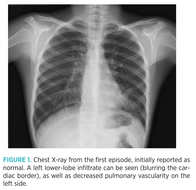
An 11-year-old boy with no relevant medical history presented to the emergency department with a 24-hour history of fever and mild dyspnea. Lung auscultation revealed decreased breath sounds on the left side and isolated expiratory wheezing. Due to the auscultatory asymmetry, a chest X-ray was performed, which was incorrectly reported as normal (Figure 1); upon later review, an infiltrate in the left lower lobe was identified. The patient was diagnosed with acute bronchitis and discharged with bronchodilator therapy.
He returned 48 hours later due to persistent fever and dyspnea. His vital signs were normal, and auscultation again demonstrated decreased breath sounds on the left side. A lung ultrasound revealed consolidation in the left lower lobe and the apex. Directed history-taking identified a choking episode with a sunflower seed shell three months earlier, coinciding with the onset of recurrent pneumonic episodes in the same lobe (three episodes were documented in the shared medical record).
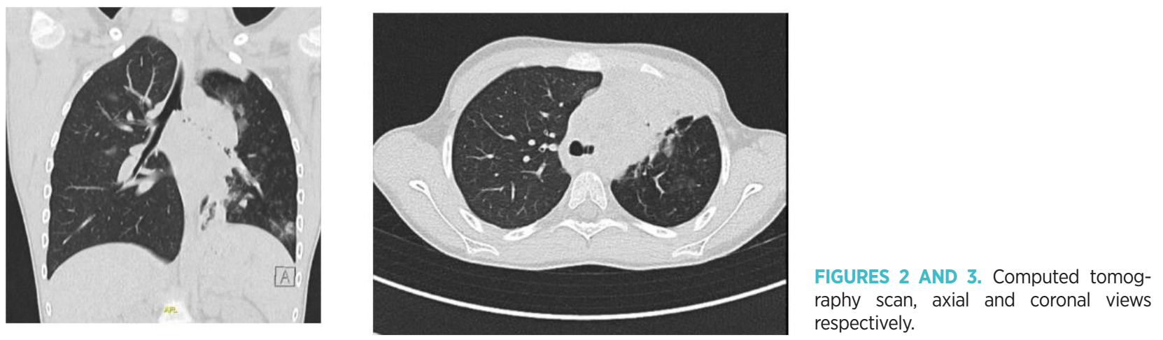
Based on these findings, the case was considered pneumonia in the context of foreign body aspiration. A chest CT scan revealed obstruction of the left main bronchus by a foreign body consistent with a seed shell, pulmonary hypovascularization, and parenchymal consolidation (Figure 2 and Figure 3).
The patient was admitted and scheduled for bronchoscopy. A flexible bronchoscopy was initially performed in the operating room and allowed visualization of the foreign body but not its extraction. A rigid bronchoscopy under general anesthesia with orotracheal intubation was then performed, successfully removing the object without complications.
Preoperative laboratory tests showed leukocytosis with neutrophilia and elevated acute phase reactants. Treatment with amoxicillin–clavulanic acid was initiated, resulting in complete clinical resolution. The patient remained asymptomatic with no further pneumonic episodes during short-term follow-up.
Radiology report:
Left lung: Extensive parenchymal consolidation predominantly affecting the lower lobe and including the pulmonary apex, compatible with an inflammatory/infectious process. Vascular hypoattenuation of the left lung parenchyma suggests hypovascularization secondary to bronchial obstruction. The left main bronchus is occupied by a hyperdense structure compatible with a foreign body, resulting in near-complete obstruction.
Right lung: Preserved parenchymal architecture with no consolidation, effusion, or visible masses. Normal ventilation and vascularization.
Tracheobronchial tree: Mediastinal shift to the right with reduced left lung volume. No other remarkable findings.
Mediastinal and pleural structures: No significant mediastinal lymphadenopathy. No evidence of pleural effusion or pneumothorax.
Conclusion:
• Foreign body in the left main bronchus with near-complete obstruction.
• Pulmonary consolidation in the left lower lobe, likely due to post-obstructive pneumonia.
• Secondary hypovascularization and hypoventilation of the left lung.
• Findings consistent with foreign body aspiration syndrome.
Discussion
This case highlights the importance of a thorough medical history and accurate interpretation of imaging studies. Recurrent pneumonia in the same lobe should prompt suspicion of an undiagnosed foreign body, especially when there is a history of choking, even if not spontaneously recalled.
A diagnostic error occurred during the interpretation of the initial chest X-ray, which already showed decreased vascularity in the left lung and a small infiltrate. However, it is important to recognize that initial diagnostic tools—particularly chest radiography—have limited sensitivity. It is estimated that 9% to 35% of chest X-rays may appear normal in confirmed aspiration cases(6,10). CT imaging, although highly sensitive (99%) and specific (97%)(6), is not always required for diagnosis and should be reserved for complex cases or when clinical uncertainty remains. In this case, the history of repeated pneumonias in the same location and the prior choking episode already suggested the diagnosis, making the CT scan not strictly necessary. CT involves a considerable radiation dose, which is especially relevant in children.
In this case, flexible bronchoscopy was initially performed because it is useful as a diagnostic tool; however, therapeutic extraction of foreign bodies in children is predominantly performed by rigid bronchoscopy. Swanson et al.(1) reported a success rate greater than 95% for rigid bronchoscopy in the removal of foreign bodies in children, while flexible bronchoscopy has shown success rates of 87% in large series(7). Flexible bronchoscopy is mainly reserved for distal foreign bodies or as a diagnostic step prior to rigid bronchoscopy, which remains the treatment of choice(8,9).
Foltran et al.(2) reported that between 40% and 50% of children with foreign bodies present with pneumonia as the initial manifestation, which can lead to diagnostic errors if the underlying cause is not investigated.
Recent studies emphasize that computed tomography has a sensitivity of 99% and specificity of 97% for detecting foreign bodies in the pediatric airway(6).
Timely extraction helps prevent complications such as bronchiectasis, atelectasis, or pulmonary fibrosis. This case also underscores the importance of maintaining a high index of suspicion and reassessing diagnostic tests when clinical progression is unfavorable.
An essential aspect in preventing complications is educating caregivers and healthcare professionals on early identification of aspiration signs. Warning signs include chronic cough, unreported prior choking episodes, persistent wheezing, and recurrent pneumonia in the same pulmonary location.
Conclusions
• Recurrent pneumonia in the same location should prompt suspicion of an airway foreign body.
• Medical history and accurate interpretation of imaging studies are essential for early diagnosis.
• CT can be useful but is not required in all cases; its use should be weighed against radiation risks in children.
• Rigid bronchoscopy remains the therapeutic gold standard for foreign body removal in children.
• Early detection and removal prevent severe pulmonary complications.
Funding
This research received no specific grant from public, commercial, or not-for-profit funding agencies.
Conflicts of Interest
The authors declare no conflict of interest.
Authors’ Contributions
The authors certify equal contributions to the scientific and intellectual content, data analysis, and writing of the manuscript.
Ethical Responsibilities
The authors confirm that informed consent was obtained from the patients involved in accordance with the ethical principles of the Declaration of Helsinki.
References
- Swanson KL, Prakash UBS, Midthun DE. Flexible bronchoscopic management of airway foreign bodies in children. Chest. 2002;121(5):1695–700. Available from: https://doi.org/10.1378/chest.121.5.1695
- Foltran F, Ballali S, Passali FM, et al. Foreign bodies in the airways: a meta-analysis of published papers. Int J Pediatr Otorhinolaryngol. 2012;76(Suppl 1):S12–9. Available from: https://doi.org/10.1016/j.ijporl.2012.02.004
- Karišik M. Foreign body aspiration and ingestion in children. Acta Clin Croat. 2023;62(Suppl 1):105–12. Available from: https://doi.org/10.20471/acc.2023.62.s1.12
- Sancho Marquina I, Aranda Conchello E, Gallán Farina IL, Martín de Vicente C. Complicaciones por cuerpo extraño intrabronquial de larga evolución. An Pediatr (Barc). 2025;102:503738. Available from: https://doi.org/10.1016/j.anpedi.2025.503738
- Fidkowski CW, Zheng H, Firth PG. The anesthetic considerations of tracheobronchial foreign bodies in children: a literature review. Anesth Analg. 2010;111(4):1016–25. Available from: https://doi.org/10.1213/ANE.0b013e3181ef3e9c
- Lai YF, Cheng SL, Lin CH. Diagnostic performance of low-dose chest CT for suspected foreign body aspiration in children. Pediatr Radiol. 2022;52(5):874–81. Available from: https://doi.org/10.1007/s00247-021-05162-2
- Alnassar M, Alotaibi S, Alazemi N, et al. Success rate and complications of flexible bronchoscopy for tracheobronchial foreign body removal in children. Int J Pediatr Otorhinolaryngol. 2022;158:111177. Available from: https://doi.org/10.1016/j.ijporl.2022.111177
- Righini CA, Morel N, Karkas A, et al. What is the best tool for the extraction of tracheobronchial foreign bodies in children? Review of 20 years of experience. Int J Pediatr Otorhinolaryngol. 2021;140:110741. Available from: https://doi.org/10.1016/j.ijporl.2020.110741
- Su D, Liu Y, Liu D, et al. Rigid vs. flexible bronchoscopy for foreign body aspiration in children: a systematic review and meta-analysis. Eur Arch Otorhinolaryngol. 2023;280(2):637–47. Available from: https://doi.org/10.1007/s00405-022-07590-y
- El Khoury P, et al. Diagnostic value of chest CT. Otolaryngol Head Neck Surg. 2024;170(1):1–12. Available from: https://doi.org/10.1002/ohn.30472
- Safia A, et al. Flexible vs rigid bronchoscopy. J Clin Med. 2024;13(18):5652. Available from: https://doi.org/10.3390/jcm13185652
- Chantzaras AP, et al. Flexible bronchoscopy meta-analysis. Acta Paediatr. 2022;111(7):1301–12. Available from: https://doi.org/10.1111/apa.16347
Explorar
Artículos
Volúmenes
Sobre la revista
Más leídos
Emergencias Pediátricas
Emergencias Pediátricas
Emergencias Pediátricas
1º Congreso Mundial de Emergencias Pediátricas
Emergencias Pediátricas
Una nueva mirada a la vía aérea pediátrica: secuencia de intubación diferida como alternativa segura en situaciones de urgencias
Emergencias Pediátricas