Artículo original
Vol. 5 Núm. 1 (English)
23 min
Evolution of infants with moderate to severe bronchiolitis treated with high-flow nasal cannulas: a comparative analysis with a historical cohort
Evolución de lactantes con bronquiolitis moderada a grave tratados con cánulas nasales de alto flujo. Análisis comparativo con una cohorte histórica
Pedro Rino*1,2, Julián Rosa1, Marianella Castagnasso1, Noelia Bonduel1, Gabriela Casais1, Ana Fustiñana1
1Prof. Dr. Juan P. Garrahan Pediatric Hospital, Autonomous City of Buenos Aires, Argentina, 2 University of Buenos Aires, Ciudad Autónoma de Buenos Aires, Argentina
Correspondencia:
Recepción:
03/05/2025
Aceptación:
19/09/2025
Publicación:
11/03/2026
Métricas
134
Visualizaciones
25
Descargas
Compartir
Compartir
Resumen (ES)
Introducción: La bronquiolitis es una enfermedad respiratoria común en lactantes y un problema sanitario global. El uso de cánulas nasales de alto flujo (CNAF) ha sido propuesto como una alternativa terapéutica para mejorar su evolución. Este estudio compara el tratamiento con oxigenoterapia estándar (O₂E) y CNAF en lactantes con bronquiolitis moderada a grave.
Material y métodos: Estudio ambispectivo y observacional realizado en un hospital de tercer nivel, con dos cohortes de lactantes menores de 2 años con bronquiolitis moderada a grave. Se compararon días de oxigenoterapia (DDO₂), días de estancia hospitalaria (DDE), admisión a unidad de cuidados intensivos pediátricos (UCIP) y necesidad de ventilación mecánica invasiva (VMI).
Resultados: Se incluyeron 427 pacientes, 223 en O2E (cohorte histórica) y 204 en CNAF (cohorte prospectiva). El grupo CNAF mostró mejoría clínica en las primeras 2 horas, pero no hubo diferencias significativas en DDO₂, DDE, admisión a UCIP ni necesidad de VMI.
Conclusión: A pesar de los beneficios iniciales en la mejora del trabajo respiratorio, los resultados finales no mostraron ventajas de la CNAF sobre O2E. Se requieren más investigaciones con metodologías robustas y criterios estandarizados para evaluar el impacto del uso de CNAF en el tratamiento de la bronquiolitis en diferentes contextos sanitarios.
Palabras clave (ES):
Resumen (EN)
Introduction: Bronchiolitis is a common respiratory disease in infants and a global health problem. The use of high-flow nasal cannulas (HFNC) has been proposed as a therapeutic alternative to improve outcomes. This study compares treatment with standard oxygen therapy (SOT) versus HFNC in infants with moderate-to-severe bronchiolitis.
Material and methods: An ambispective, observational study was conducted in a tertiary care hospital, involving two cohorts of infants under 2 years of age with moderate-to-severe bronchiolitis. Comparisons were made regarding duration of oxygen therapy (DOOT), length of hospital stay (LOS), admission to the pediatric intensive care unit (PICU), and the need for invasive mechanical ventilation (IMV).
Results: A total of 427 patients were included: 223 in the SOT group (historical cohort) and 204 in the HFNC group (prospective cohort). The HFNC group showed clinical improvement within the first 2 hours; however, there were no significant differences in DOOT, LOS, PICU admission, or the need for IMV.
Conclusion: Despite initial benefits in improving the work of breathing, the final results showed no advantages of HFNC over SOT. Further research with robust methodologies and standardized criteria is required to evaluate the impact of HFNC use in the treatment of bronchiolitis across different healthcare settings.
Palabras clave (EN):
Introduction
Bronchiolitis is a global health problem in infants and one of the ten most common diagnoses in pediatric critical illness(1,2). Respiratory syncytial virus (RSV) is the most frequent cause, with more than 30,000,000 cases, 3,000,000 hospitalizations, and nearly 200,000 deaths per year, mainly in low-resource countries(3-5). Between 1% and 5% of affected children require hospitalization, and up to 15% are admitted to pediatric intensive care units (PICUs), with mechanical ventilation (MV) rates ranging from 5% to 12%(5-11).
RSV infections show epidemic peaks during cold and humid months(12), increasing healthcare demand and potentially saturating pediatric emergency departments (PEDs), inpatient wards, and PICUs.
Multiple strategies have been proposed to reduce its health impact: preventive measures, such as the promotion of breastfeeding(13) and smoke-free environments(14) and RSV immunization(15), and therapeutic options, such as inhaled ꞵ2 agonists(16,17), inhaled ipratropium bromide(18), nebulized adrenaline(19,20), inhaled and systemic corticosteroids(21), nebulized hypertonic saline solution(19,20,22), nebulized and intravenous magnesium sulfate(23), and nebulized helium-oxygen(24), although none have solid evidence for reducing length of stay (LOS), PICU admission, or invasive mechanical ventilation (IMV).
The complexity of its analysis is further increased by the variability in definitions(25-28), the variety of severity classification scales(29-31), and the existence of “clinical phenotypes” in infants based on age, history of atopy, etiology, and pathophysiology, which suggests that some cases may correspond to the first episode of asthma(32,33).
Treatment is based on hydration, nutrition, secretion aspiration, oxygen therapy, and ventilatory support according to the patient’s severity.
In recent decades, humidified and heated high-flow nasal cannulas (HFNC) have been used for respiratory failure, high oxygen (O2) requirements, upper airway obstruction, post-extubation laryngitis, heart failure, VM weaning, apnea, asthma, and bronchiolitis(34).
This system is safe, well-tolerated, and does not interfere with feeding. It allows for the use of high flows with high O₂ concentrations. It offers benefits such as decreased upper airway resistance, improved ciliary activity, reduced secretion viscosity and inflammation, reduction of bronchoconstriction induced by cold and dry air, decreased nasopharyngeal dead space, and a certain degree of continuous positive airway pressure (CPAP) that would contribute to improving ventilation and reducing the work of breathing(35-40).
It has been suggested that HFNC therapy could decrease respiratory distress, the need for IMV, DOOT, and LOS, in addition to adverse effects and costs(41-45). However, the evidence remains insufficient, and most studies come from countries with greater economic and healthcare resources.
The objective of this work is to describe the evolution of the first cohort of patients in our center with moderate to severe bronchiolitis treated with HFNC and compare it with a historical cohort treated with SOT.
Materials and Methods
An ambispective, comparative, and observational study of two cohorts (prospective and historical) of children under 2 years of age with a discharge diagnosis of bronchiolitis was conducted. These patients consulted the PED of a tertiary care hospital in Buenos Aires, Argentina.
Infants between 1 and 24 months of age, born at term, presenting with the first or second episode of moderate to severe bronchiolitis, defined by a modified Tal Scale score (Tal) ≥ 7, according to the criteria established by the Argentinian Society of Pediatrics at that time, were included(4). Patients with imminent acute respiratory failure, altered consciousness (Glasgow Coma Scale score < 15), hemodynamic instability, apneas, choanal atresia or upper respiratory obstruction, severe craniofacial malformations, facial trauma, nasopharyngeal surgery, pneumothorax, suspected or confirmed foreign body aspiration, pre-existing lung disease, congenital heart disease, and other chronic conditions were excluded.
Based on the studies by Kepreotes et al.(44) and Franklin et al.(45), which reported a decrease in the need for care escalation of 58% and 48%, respectively, we decided to use this background to calculate the sample size. Assuming that 15% of infants with bronchiolitis require PICU admission, we proposed a reduction of this percentage to 6% with the use of HFNC. Based on this hypothesis, a sample size of 204 patients per group was calculated, with a 95% confidence level and 80% power.
To reach the sample size, the prospective cohort included a consecutive sample of patients between July 2017 and September 2018. For the historical cohort, data obtained from the Statistics Office corresponding to patients hospitalized between June 2016 and May 2017 who were discharged with a diagnosis of bronchiolitis were used. From the 1828 records obtained, a randomized sampling was performed to reach the calculated sample size, leaving 204 electronic records to be analyzed. Since a sample with a higher proportion of severe bronchiolitis (Tal ≥ 9) was obtained in the HFNC cohort, it was decided to include more infants with severe bronchiolitis in the historical cohort to balance this difference.
In both cohorts, the treatment was based on institutional protocols, and the only variation was the incorporation of HFNC in the treatment of moderate to severe bronchiolitis (Tal ≥ 7) in the prospective cohort.
HFNC was used with a flow of 2 L/kg/min through Optiflow® cannulas and an MR 810/850 heater-humidifier (Fisher & Paykel®). An initial fraction of inspired oxygen (FiO2) of 0.6 was configured using an Airvo2® device, a gas blender, or ‘Y’-connected tubing to the corresponding O₂ and compressed air flow meters. FiO2 was optimized according to clinical evolution after the first 4 hours of observation. Patients with a good response remained in the SUP or general wards; those without improvement were transferred to the PICU.
In the historical cohort, O2 was administered with conventional cannulas (maximum flow 2 L/min), Venturi masks, or non-rebreather masks, without humidification or heating.
Demographic variables and breastfeeding were recorded in both populations. Variables related to clinical presentation, pulse oximetry on admission, and the result of the virological study of the nasopharyngeal secretions aspirate were also included.
Among the primary outcomes, evolution was assessed at 2, 72, and > 72 hours, defining good treatment response as a Tal score ≤ 6. In the retrospective cohort, these variables were obtained by analyzing vital signs charts and clinical records available in the electronic medical history. Likewise, variables such as DOOT, LOS, PICU admission, and need for IMV were analyzed.
Other variables recorded included the use of non-invasive mechanical ventilation (NIMV), the incidence of cardiorespiratory arrest (CRA), respiratory arrest (RA), 28-day mortality, and complications associated with HFNC, such as air leaks (pneumothorax, pneumomediastinum, pneumopericardium, or pulmonary interstitial emphysema), nasal injury (erythema, erosion, ulcer), and gastric distension.
Categorical variables were described with frequencies and continuous variables with median and interquartile ranges (IQR), and Chi-square Test and Mann-Whitney U test were applied for their comparisons, respectively.
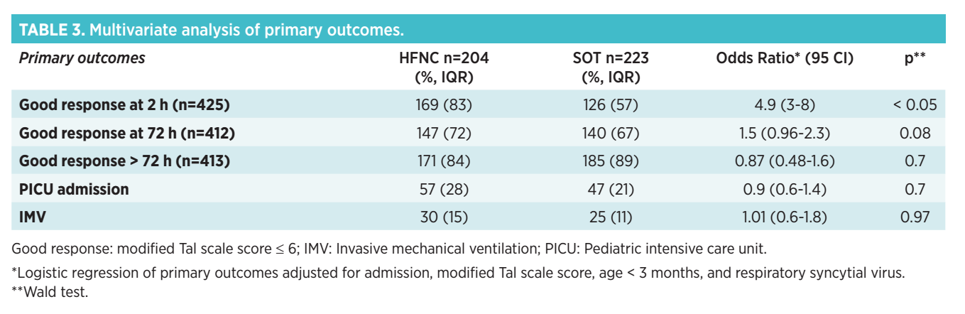
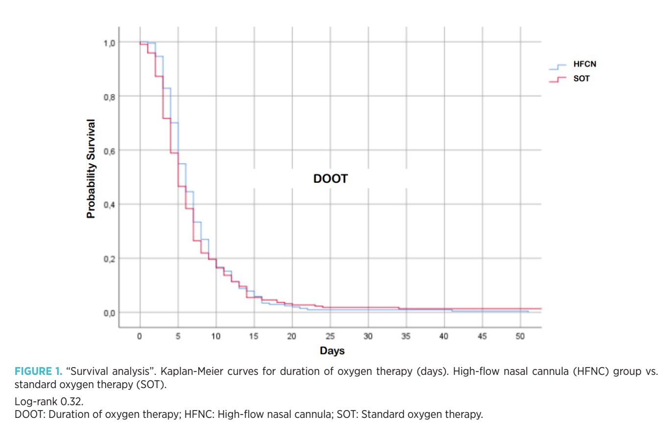
For dichotomous primary outcomes (good response at 2, 72, and > 72 hours, PICU admission, and IMV), multivariate logistic regression models were performed to adjust each one for confounders. The variables included in the adjustment were those with p-values < 0.2 in the univariate analysis. Odds ratios and their 95% CIs were reported. For time-to-event outcomes (DOOT and LOS), survival curves were plotted using the Kaplan-Meier method, and the log-rank test was reported. A p-value < 0.05 was considered statistically significant.
RedCap® was used as computer support for data entry, and SPSS® version 24 for statistical analysis.
The protocol was approved by the Research and Ethics Committees, without requiring informed consent, as the treatment corresponded to the institutional protocol.
Results
After performing randomized sampling of the historical cohort and selecting 204 patients out of 1828, it was observed that both populations presented similar demographic characteristics, except for a higher proportion of children with severe bronchiolitis (Tal score ≥ 9) in the HFNC cohort. To correct this difference, it was decided to include more infants with severe bronchiolitis in the historical cohort. However, even after adding all severe bronchiolitis cases recorded during the study period for the SOT group, the number remained lower.
Finally, 427 children were included: 223 in the SOT population and 204 in the HFNC population (Table 1).
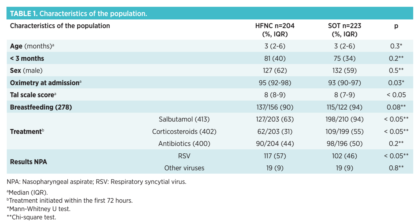
The results of the analyzed variables are shown in Table 2.
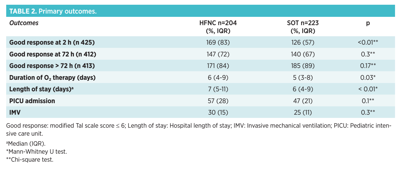
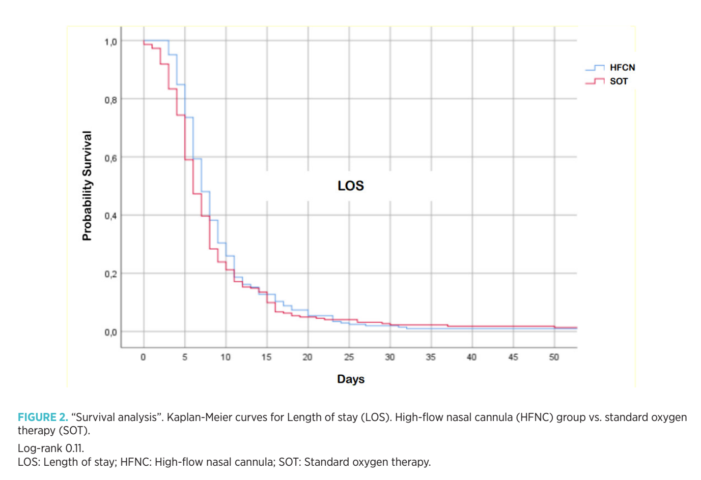
A better immediate response (improvement in Tal score) was observed in the HFNC cohort compared to the SOT cohort. Both DOOT and LOS were longer in the HFNC group. However, as shown in the survival curves in Graphs 1 and 2, these differences were not significant (log-rank test for duration of oxygen therapy: 0.32; log-rank test for LOS: 0.11).
There were no significant differences between the two groups regarding the use of NIV [HFNC group 29 (14%) vs. SOT group 27 (12%), p = 0.5], PICU admission, and requirement for IMV.
When multivariate analysis was performed, a better response within 2 hours of treatment was sustained in the HFNC group, but there were no significant differences in PICU admission and need for IMV (Table 3).
Neither mortality nor cardiorespiratory arrest events were observed in any group. Only one respiratory arrest occurred in the HFNC cohort. No complications associated with HFNC were recorded.
Discussion
There is experience with HFCN in Latin America; however, there are few publications comparing its use with SOT. The available studies suggest a possible reduction in LOS and MV(41-43).
The choice of evaluation intervals in this study (at 2, 72, and > 72 hours) responded to clinical and methodological criteria, to distinguish the immediate, mediate, and later response where other factors inherent to the hospitalization may influence the outcome.
Regarding the characteristics of the studied population, it should be considered that the higher proportion of more severe patients in the HFCN group could be due, in part, to the limited availability of this resource at the beginning of the study. This may have generated a selection bias towards more severe cases, under the premise that HFCN would offer greater benefits compared to SOT.
Despite having a higher initial proportion of severe patients, the HFCN cohort had similar outcomes for LOS, DOOT, and MV to the SOT cohort. This finding, which could suggest a benefit, must be interpreted with great caution and confirmed in prospective studies with robust methodologies and standardized criteria.
Significant differences were found in some administered treatments between both populations. This could be explained by greater adherence to recommendations that discourage inhaled β2 agonists, corticosteroids, and antibiotics in the treatment of bronchiolitis.
In 2017, Kepreotes et al. published a prospective, randomized, controlled study in Australia with 202 children under 2 years of age with moderate bronchiolitis(44). A year later, Franklin et al. reported a similar, multicenter study in Australia and New Zealand, with approximately 1500 children under 1 year of age with different degrees of severity(45).
Both studies compared HFCN vs. conventional low-flow O₂ nasal cannulas, observing an increase in the escalation of care and treatment failure in the second group, according to criteria based on vital signs, early warning tools, and medical judgment. Although these results seemed quite promising, no significant differences were found in secondary outcomes such as DOOT, LOS, PICU admission, and intubation.
Durand et al. published a prospective, randomized, multicenter study in France in 2020, where they did not observe significant differences regarding the need to escalate ventilatory support. The use of HFNC also did not reduce LOS or PICU admission(46).
O’Brien et al. reported in 2022 an increase in the use of HFNC and even greater LOS, PICU admissions, NIV, and IMV in populations that used this therapy(47).
In 2024, Armarego et al. published a systematic review that included 11 randomized or quasi-randomized controlled trials and concluded modest reductions in DOOT, LOS, and need for treatment escalation (criteria varied by study) with a low to moderate level of evidence(48).
Antilici et al., in a retrospective quasi-experimental analysis spanning 2021-2022 and 2022-2023, also mentioned that HFNC did not reduce DOOT or LOS, nor were there significant differences in treatment failure compared to SOT (49).
In a cohort study with historical control, Gutiérrez Moreno et al. also found no association with lower DOOT, LOS, or transfer to PICU (50).
Similarly, our analysis also showed no differences in LOSO2, LOS, PICU admission, and IMV between both populations.
Considering the study results, it has been discussed whether the increased use of HFNC is associated with increased LOS, PICU admissions, and MV.
On the one hand, in a retrospective analysis of a tertiary center, Tirelli et al. did not observe a greater tendency for PICU admissions or greater LOS(51). On the other hand, multicenter studies by O’Brien et al.(47) and Pelletier et al.(52) described an increase in the use of HFNC accompanied by an increase in PICU and NIMV admissions, but without a concomitant decrease in IMV.
In our series, there is a certain trend toward an increase in PICU admissions in the HFNC group.
As described by Kepreotes et al.(44), Durand et al. (46), and Armarego et al.(48), we also verified lower work of breathing in the HFNC group during the first hours after device installation. It is worth asking whether HFNC could favor an increase in initial care levels, contributing to early improvement in the work of breathing.
HFNC is a safe and simple therapy to implement. In our research, as in those of Kepreotes et al.(44) and Franklin et al.(45), no adverse events associated with its use were observed.
Furthermore, there was no mortality, and only one RA in the HFNC group was recorded, which cannot necessarily be associated with its use.
In turn, cost-effectiveness analyses have suggested that the use of HFNC as first-line treatment versus SO2 in cases of bronchiolitis and hypoxemic acute respiratory failure is not an efficient option(53-55).
Our research presented both strengths and limitations.
This is one of the few publications in Latin America that compares the evolution of infants with moderate to severe bronchiolitis treated with SOT vs. HFNC, contributing to knowledge in a regionally understudied context. From an epidemiological perspective, it provides evidence both in Latin America and globally.
The detailed definition of inclusion and exclusion criteria improves the homogeneity of the cohorts and ensures that the results are applicable only to patients with moderate to severe bronchiolitis.
Having standardized institutional treatment protocols in both cohorts allowed for a comparative evaluation of the effectiveness of the two therapeutic approaches.
The study compared a prospective cohort with a randomized retrospective sample, but differences were found between the two, particularly a higher proportion of patients with severe bronchiolitis in the HFNC cohort. To mitigate this disparity, it was decided to include all patients with a Tal score ≥ 9 in the historical cohort; the difference persisted, but multivariate analysis attempted to lessen those confounding factors.
Incomplete records in the SOT cohort, especially those related to personal and family history (breastfeeding, asthma, eczema, and smoking), could have compromised the precision of the findings. Something similar occurred with the information on the use of inhaled β₂ agonists, corticosteroids, and antibiotics (e.g., dose, duration of treatment).
The ambispective design may introduce selection bias, but this was mitigated by taking a randomized sample from the period immediately preceding the implementation of HFNC as a treatment protocol.
We do not believe that the variable availability of different types of HFNC therapy significantly affected our results. The verification of flows and FiO2 provided by the devices were equivalent.
The tools used to define the escalation of therapeutic support may vary between healthcare centers and be conditioned by the availability of resources, which could even influence relevant clinical decisions, such as admission to the PICU. In this context, an objective datum such as the need for IMV is more representative for evaluating clinical outcomes.
By focusing on a local population, the findings acquire relevance for regional clinical practice, although their extrapolation to other populations must be done cautiously.
Although the study presents limitations inherent to its design, the results are consistent with those reported internationally.
Finally, as this was a single-center study, the generalization of its conclusions is limited.
Conclusion
Although there was an improvement in the work of breathing during the first hours of HFNC therapy, in our study we did not observe a decrease in DOOT, DDE, PICU admission, and IMV in infants with moderate to severe bronchiolitis. More studies with good methodological quality and standardized definitions and criteria are needed to establish a clear recommendation on its use and adaptation to different health and socioeconomic realities.
Acknowledgments
To Dr. Susana Rodríguez and all the staff of the Emergency, Intermediate and Moderate Care, and Intensive Care Departments of the "Prof. Dr. Juan P. Garrahan" Pediatrics Hospital.
Funding
No funding was received for the research, authorship, and/or publication of this article.
Author contributions
Pedro Rino: Conceptualization, Methodology, Formal analysis, Writing – original draft, Review and editing, Supervision, Validation, Funding, Project administration.
Ana Fustiñana: Methodology, Formal analysis, Data collection, Data curation, Writing – Review and editing.
Julián Rosa: Investigation, Data collection, Writing – review and editing.
Marianela Castagnasso: Investigation, Data collection, Writing – review and editing.
Noelia Bonduel: Investigation, Data collection, Writing – review and editing.
Gabriela Casais: Investigation, Data collection, Writing – review and editing.
All authors have read and accepted the final version of the manuscript.
Conflicts of interest
The authors declare that there are no commercial or financial conflicts of interest for this research.
Use of artificial intelligence tools
The authors declare that no artificial intelligence tool (such as ChatGPT, Copilot, Gemini, or others) has been used in the writing, analysis, or review of this article.
References
- Kyu HH, Pinho C, Wagner JA, Brown JC, Bertozzi-Villa A, Charlson FJ et al. Global and national burden of diseases and injuries among children and adolescents between 1990 and 2013: findings from the Global Burden of Disease 2013 Study. JAMA Pediatr. 2016; 170: 267-287.
- Kortz TB, Holloway A, Agulnik A, He D, Rivera SG, Abbas Q et al; Global PARITY Investigators; Global Health Subgroup of the Pediatric Acute Lung Injury and Sepsis Investigators (PALISI) Network. Prevalence, aetiology, and hospital outcomes of paediatric acute critical illness in resource-constrained settings (Global PARITY): a multicentre, international, point prevalence and prospective cohort study. Lancet Glob Health. 2025; 13(2): e212-e221.
- Nair H, Nokes DJ, Gessner BD, Dherani M, Madhi SA, Singleton RJ et al. Global burden of acute lower respiratory infections due to respiratory syncytial virus in young children: a systematic review and meta-analysis. Lancet. 2010; 375: 1545-1555.
- Comité Nacional de Neumonología. Subcomisión de Epidemiología. Comité Nacional de Infectología. Comité Nacional de Medicina Interna. Recomendaciones para el diagnóstico y tratamiento de las infecciones respiratorias agudas bajas en menores de 2 años. Arch Argent Pediatr. 2006; 104: 159-176.
- Oñoro G, Pérez Suárez E, Iglesias Bouzas MI, Serrano A, Martínez De Azagra A, García-Teresaet MA al. Bronquiolitis grave. Cambios epidemiológicos y de soporte respiratorio. An Pediatr (Barc). 2011; 74(6): 371-376.
- Golubicki A, Gómez Traverso R. Observatorio de Enfermedades Respiratorias Agudas Bajas en Unidades de Internación Pediátricas. Ministerio de Salud de la Ciudad de Buenos Aires. 2015.
- Tian J, Wang XY, Zhang LL, Liu MJ, Ai JH, Feng GS, et al. Clinical epidemiology and disease burden of bronchiolitis in hospitalized children in China: a national cross-sectional study. World J Pediatr. 2023; 19(9): 851-863.
- Heppe Montero M, Gil-Prieto R, Walter S, Aleixandre Blanquer F, Gil De Miguel Á. Burden of severe bronchiolitis in children up to 2 years of age in Spain from 2012 to 2017. Hum Vaccin Immunother. 2022; 18(1):1883379.
- Barbieri E, Cavagnis S, Scamarcia A, Cantarutti L, Bertizzolo L, Bangert M et al. Assessing the burden of bronchiolitis and lower respiratory tract infections in children ≤ 24 months of age in Italy, 2012-2019. Front Pediatr. 2023; 11:1143735.
- Mahant S, Parkin PC, Thavam T, Imsirovic H, Tuna M, Knight B et al; Canadian Paediatric Inpatient Research Network (PIRN). Rates in Bronchiolitis Hospitalization, Intensive Care Unit Use, Mortality, and Costs From 2004 to 2018. JAMA Pediatr. 2022; 176(3): 270-279.
- Camporesi A, Yock-Corrales A, Gomez-Vargas J, Roland D, Gonzalez M, Barreiro S et al. Management and outcomes of bronchiolitis in Italy and Latin America: a multi-center, prospective, observational study. Eur J Pediatr. 2024; 183(6): 2733-2742.
- Haynes AK, Manangan AP, Iwane MK, Sturm-Ramirez K, Homaira N, Brooks WA et al. Respiratory syncytial virus circulation in seven countries with Global Disease Detection Regional Centers. J Infect Dis. 2013; 208 (suppl 3): S246-54.
- Mineva G, Philip R. Impact of breastfeeding on the incidence and severity of respiratory syncytial virus bronchiolitis in infants: systematic review. Rural Remote Health. 2023; 23(1): 8088.
- Turner S, Mackay D, Dick S, Semple S, Pell JP. Associations between a smoke-free homes intervention and childhood admissions to hospital in Scotland: an interrupted time-series analysis of whole-population data. Lancet Public Health. 2020; 5(9): e493-e500.
- Vázquez-López P, Rivas-García A, Luaces-Cubells C, Perez-Gonzalez E, Ales-Palmer ML, Cahis-Vela N et al. Changes in Care in Spanish Pediatric Emergency Departments After the First Immunization With Nirsevimab. Pediatr Emerg Care. 2025; 41(5): 365-371.
- Gómez C, Alarcón G, Cifuentes L. Beta-2 agonists for the treatment of bronchiolitis. Medwave. 2020; 20(8): e7945.
- Nino G, Rodríguez-Martínez CE, Castro-Rodríguez JA. The use of β2-adrenoreceptor agonists in viral bronchiolitis: scientific rationale beyond evidence-based guidelines. ERJ Open Res. 2020; 6(4): 00135-2020.
- Everard ML, Bara A, Kurian M, Elliott TM, Ducharme F, Mayowe V. Anticholinergic drugs for wheeze in children under the age of two years. Cochrane Database Syst Rev. 2005; 2005(3): CD001279.
- Pereira RA, Oliveira de Almeida V, Zambrano M, Zhang L, Amantéa SL. Effects of nebulized epinephrine in association with hypertonic saline for infants with acute bronchiolitis: A systematic review and meta-analysis. Health Sci Rep. 2022; 5(3): e598.
- Elliott SA, Gaudet LA, Fernandes RM, Vandermeer B, Freedman SB, Johnson DW et al. Comparative Efficacy of Bronchiolitis Interventions in Acute Care: A Network Meta-analysis. Pediatrics. 2021; 147(5): e2020040816.
- Fernandes RM, Bialy LM, Vandermeer B, Tjosvold L, Plint AC, Patel H et al. Glucocorticoids for acute viral bronchiolitis in infants and young children. Cochrane Database Syst Rev. 2013; 2013(6): CD004878.
- Heikkilä P, Renko M, Korppi M. Hypertonic saline inhalations in bronchiolitis-A cumulative meta-analysis. Pediatr Pulmonol. 2018; 53(2): 233-242.
- Chandelia S, Kumar D, Chadha N, Jaiswal N. Magnesium sulphate for treating acute bronchiolitis in children up to two years of age. Cochrane Database Syst Rev. 2020; 12(12):CD012965.
- Kuitunen I, Kiviranta P, Sankilampi U, Salmi H, Renko M. Helium-oxygen in bronchiolitis-A systematic review and meta-analysis. Pediatr Pulmonol. 2022; 57(6): 1380-1391.
- Yanes Macías JC, Fonseca Hernández M, García Rodríguez I, Llul Tombo C, Tio González D, Díaz Ceballos JC. Atención al niño con bronquiolitis: consideraciones clínico-terapéuticas generales. MediSur Cienfuegos. 2022; 20(2): 175-182.
- Benito Fernández J, Paniagua Calzón N. Diagnóstico y tratamiento de la bronquiolitis aguda en Urgencias. Protoc diagn ter pediatr. 2020; 1: 63-73. SEUP. [Consulta: 7 de febrero de 2025]. Available from: https://www.aeped.es/protocolos/y https://www.aeped.es/sites/default/files/documentos/05_bronquiolitis.pdf.
- National Institute for Health and Care Excellence. Bronchiolitis in children: diagnosis and management. NICE guideline. [Consulta: 3 de marzo de 2025]. Available from: https://www.nice.org.uk/guidance/ng9/resources/bronchiolitis-in-children-diagnosis-and-management-pdf-51048523717.
- Justice NA, Le JK. Bronchiolitis. [Consulta: 3 de marzo de 2025]. Available from: https://www.ncbi.nlm.nih.gov/books/NBK441959
- Sheikh Z, Potter E, Li Y, Cohen RA, Dos Santos G, Bont L et al; PROMISE Investigators. Validity of Clinical Severity Scores for Respiratory Syncytial Virus: A Systematic Review. J Infect Dis. 2024; 229(Supplement_1): S8-S17.
- Granda E, Urbano M, Andrés P, Corchete M, Cano A, Velasco R. Comparison of severity scales for acute bronchiolitis in real clinical practice. Eur J Pediatr. 2023; 182(4): 1619-1626.
- Freire G, Kuppermann N, Zemek R, Plint AC, Babl FE, Dalziel SR et al; Pediatric Emergency Research Networks (PERN). Predicting Escalated Care in Infants With Bronchiolitis. Pediatrics. 2018; 142(3): e20174253. Erratum in: Pediatrics. 2019; 143(2): e20183404.
- Bottau P, Liotti L, Laderchi E, Palpacelli A, Calamelli E, Colombo C et al. Something Is Changing in Viral Infant Bronchiolitis Approach. Front Pediatr. 2022; 10:865977.
- Walsh PS, Wendt WJ, Lipshaw MJ. Asthmalitis? Diagnostic Variability of Asthma and Bronchiolitis in Children < 24 Months. Hosp Pediatr. 2024; 14(1): 59-66.
- Prego Petit J. Oxigenoterapia por cánulas nasales de alto flujo. Emerg Pediatr. 2023; 2(1): 27-32.
- Mikalsen IB, Davis P, Øymar K. High flow nasal cannula in children: a literature review. Scand J Trauma Resusc Emerg Med. 2016; 24: 93.
- Masclans JR, Peréz-Terán P, Roca O. Papel de la oxigenoterapia de alto flujo en la insuficiencia respiratoria aguda. Med Intensiva. 2015; 39: 505-515.
- Kawaguchi A, Yasui Y, deCaen A, Garros D. The Clinical Impact of Heated Humidified High-Flow Nasal Cannula on Pediatric Respiratory Distress. Pediatr Crit Care Med. 2017; 18: 112-119.
- Weiler T, Kamerkar A, Hotz J, Ross PA, Newth CJL, Khemani RG. The Relationship between High Flow Nasal Cannula Flow Rate and Effort of Breathing in Children. J Pediatr. 2017; 189: 66-71.e3.
- Pham TM, O’Malley L, Mayfield S, Martin S, Schibler A. The effect of high flow nasal cannula therapy on the work of breathing in infants with bronchiolitis. Pediatr Pulmonol. 2015; 50(7): 713-720.
- Urbano Villaescusa J, Mencía Bartolomé S, Cidoncha Escobar E, López-Herce Cid J, Santiago Lozano MJ, Carrillo Álvarez A. Experiencia con la oxigenoterapia de alto flujo en cánulas nasales en niños. An Pediatr. 2008; 68: 4-8.
- Morosini F, Dall´Orso P, Alegretti M, Alonso B, Rocha S, Cedres A et al. Impacto de la implementación de oxigenoterapia de alto flujo en el manejo de la insuficiencia respiratoria por infecciones respiratorias agudas bajas en un departamento de emergencia pediátrica. Arch Pediatr Urug. 2016; 87(2): 87-94.
- Monteverde E, Fernández A, Ferrero F, Barbaro C, De Lillo L, Lavitola M et al. High-flow nasal cannula oxygen therapy in infants with acute lower respiratory tract infection. An experience in hospitals of the City of Buenos Aires. Arch Argent Pediatr. 2019; 117(5): 286-293.
- Hernández Dinza P, Villalón Artires P, Sánchez Mancebo K, Alexander Pérez A, Rodríguez Aguirre Y. Comparación de la oxigenación de alto flujo con la oxigenoterapia convencional en niños con bronquiolitis. Multimed. 2022; 26(1).
- Kepreotes E, Whitehead B, Attia J, Oldmeadow C, Collison A, Searles A et al. High-flow warm humidified oxygen versus standard low-flow nasal cannula oxygen for moderate bronchiolitis (HFWHO RCT): an open, phase 4, randomised controlled trial. Lancet. 2017; 389(10072): 930-939.
- Franklin D, Babl FE, Schlapbach LJ, Oakley E, Craig S, Neutze J et al. A Randomized Trial of High-Flow Oxygen Therapy in Infants with Bronchiolitis. N Engl J Med. 2018; 378(12): 1121-1131.
- Durand P, Guiddir T, Kyheng C, Blanc F, Vignaud O, Epaud R et al; Bronchopti study group. A randomised trial of high-flow nasal cannula in infants with moderate bronchiolitis. Eur Respir J. 2020; 56(1): 1901926.
- O’Brien S, Haskell L, Schembri R, Gill FJ, Wilson S, Borland ML et al.; Paediatric Research in Emergency Departments International Collaborative (PREDICT) network, Australasia. Prevalence of high flow nasal cannula therapy use for management of infants with bronchiolitis in Australia and New Zealand. J Paediatr Child Health. 2022; 58(12): 2230-2235.
- Armarego M, Forde H, Wills K, Beggs SA. High-flow nasal cannula therapy for infants with bronchiolitis. Cochrane Database Syst Rev. 2024; 3(3): CD009609.
- Antilici L, Vittucci AC, Cristaldi S, Musolino AMC, Pisani M, Rotondi Aufiero L et al. High-flow nasal cannulas versus standard oxygen therapy for moderate bronchiolitis: a quasi-experimental analysis. Pediatr Pulmonol. 2025; 60(1): e27358.
- Gutiérrez Moreno M, Barajas Sánchez V, Gil Rivas T, Hernández González N, Marugán Isabel V, Ochoa-Sangrador C. Efectividad de la oxigenoterapia de alto flujo en hospitales de segundo nivel en bronquiolitis. An Pediatr (Engl Ed). 2022; 96(6): 485-491.
- Tirelli F, Todeschini Premuda M, Francaviglia G, Frigo AC, Baraldi E, Da Dalt L et al. A two-tiered high-flow nasal cannula approach does not increase intensive care utilization and hospital length of stay in bronchiolitis. Eur J Pediatr. 2024; 183(9): 4133-4137.
- Pelletier JH, Maholtz DE, Hanson CM, Nofziger RA, Forbes ML, Besunder JB et al. Respiratory Support Practices for Bronchiolitis in the Pediatric Intensive Care Unit. JAMA Netw Open. 2024; 7(5): e2410746.
- Bayón Yusta JC, Gutiérrez Iglesias A, Galnares-Cordero L. Eficiencia de la oxigenoterapia de alto flujo para el tratamiento de dificultades respiratorias en pediatría. Ministerio de Sanidad. Servicio de Evaluación de Tecnologías Sanitarias del País Vasco; 2021. Informes de Evaluación de Tecnologías Sanitarias: OSTEBA [Consulta: 27 de marzo de 2025]. Available from: https://www.euskadi.eus/contenidos/informacion/osteba_publicacion/eu_def/adjuntos/Eficiencia_de_la_oxigenoterapia_de_alto_flujo.pdf
- Gannon B, Franklin D, Vo V, Babl FE, Schibler A. Cost-effectiveness of nasal high-flow in children with acute hypoxaemic respiratory failure. J Paediatr Child Health. 2024; 60(6): 229-239.
- Winer JC, Mertens EO, Bettin K, McCoy E, Arnold SR. Variation and Outcomes of Hospital-Level High-Flow Nasal Cannula Usage Outside of Intensive Care. Hosp Pediatr. 2022; 12(12):1087-1093.
Explorar
Artículos
Volúmenes
Sobre la revista
Más leídos
Emergencias Pediátricas
Emergencias Pediátricas
Emergencias Pediátricas
1º Congreso Mundial de Emergencias Pediátricas
Emergencias Pediátricas
Una nueva mirada a la vía aérea pediátrica: secuencia de intubación diferida como alternativa segura en situaciones de urgencias
Emergencias Pediátricas