Caso clínico
Vol. 5 Núm. 1 (English)
10 min
Erythema nodosum secondary to acute Epstein-Barr virus infection. A case report
Eritema nodoso secundario a infección aguda por el virus de Epstein-Barr. A propósito de un caso
Javier Perez-Lopez*, Maria Angeles Garcia-Herrero, Carmen Moreno-Solera, Elena Marquez-Caceres
Department of Pediatrics, Príncipe de Asturias University Hospital, Alcalá de Henares, Madrid, Spain
Correspondencia:
Recepción:
15/09/2025
Aceptación:
23/01/2026
Publicación:
11/03/2026
Métricas
172
Visualizaciones
16
Descargas
Compartir
Compartir
Resumen (ES)
El eritema nodoso (EN) es una paniculitis septal caracterizada por nódulos eritematosos y dolorosos que suelen localizarse de manera simétrica en superficies extensoras, principalmente en los miembros inferiores. Esta patología presenta una etiología multifactorial, generalmente relacionada con una reacción inmunológica de hipersensibilidad. En pediatría, el eritema nodoso es hoy en día poco frecuente, pero clínicamente relevante, ya que su diagnóstico requiere una evaluación minuciosa para identificar y tratar la causa subyacente.
Presentamos el caso de un paciente pediátrico diagnosticado de eritema nodoso en el contexto de una primoinfección por virus de Epstein-Barr (VEB), con el objetivo de destacar la importancia del reconocimiento de esta entidad en el ámbito de Urgencias Pediátricas, así como de un abordaje diagnóstico integral y multidisciplinar que permita identificar la etiología subyacente y evitar tratamientos innecesarios.
Palabras clave (ES):
Resumen (EN)
Erythema nodosum (EN) is a septal panniculitis characterized by erythematous and painful nodules that are usually symmetrically distributed on extensor surfaces, mainly on the lower limbs. This condition has a multifactorial etiology, generally related to an immunological hypersensitivity reaction. In pediatrics, erythema nodosum is currently uncommon but clinically relevant, as its diagnosis requires a thorough evaluation to identify and treat the underlying cause.
We present the case of a pediatric patient diagnosed with erythema nodosum in the context of a primary Epstein–Barr virus infection (EBV), with the aim of highlighting the importance of recognizing this entity in the Pediatric Emergency Department, as well as the need for a comprehensive and multidisciplinary diagnostic approach that allows identification of the underlying etiology and avoidance of unnecessary treatments.
Palabras clave (EN):
Introduction
Erythema nodosum (EN) is an inflammatory septal panniculitis without associated vasculitis, characterized by the abrupt onset of erythematous, warm, painful, and generally symmetric nodules on extensor surfaces, mainly of the lower limbs(1-2). These lesions can be triggered by a wide variety of stimuli. The pathophysiology is related to a hypersensitivity reaction mediated by immune complexes that leads to inflammation of the subcutaneous tissue(3).
This is an uncommon entity in the pediatric population, being more frequent in adolescence, with a low incidence in children under 2 years old(4-5). Diagnosis is mainly clinical, and complementary studies aim to identify the underlying etiology.
Treatment is usually symptomatic, with spontaneous resolution of the lesions, without ulceration, scarring, or atrophy, and recurrence is infrequent(3-5).
We present the case of a pediatric patient with erythema nodosum associated with Epstein-Barr virus (EBV) infection, highlighting the importance of a comprehensive diagnostic and therapeutic approach in these cases.
Case Report
An 8-year-old male patient, previously healthy, was referred to the Pediatric Emergency Department from primary care due to the appearance of multiple cutaneous nodules on both legs, evolving over one week. He was afebrile, without respiratory or gastrointestinal symptoms, in good general condition, with adequate intake, and normal diuresis and bowel transit. There was no history of trauma. He had a history of pharyngoamygdalitis treated with penicillin for 10 days three weeks before the current episode, without microbiological testing at that time.
On physical examination, multiple indurated, warm, and painful nodules were observed, with a smooth surface and an erythematous-violaceous color, measuring between 2 and 3 cm, distributed on the anterior aspect of both pretibial areas, without involvement in other areas. The rest of the examination was normal (Figures 1 and 2).
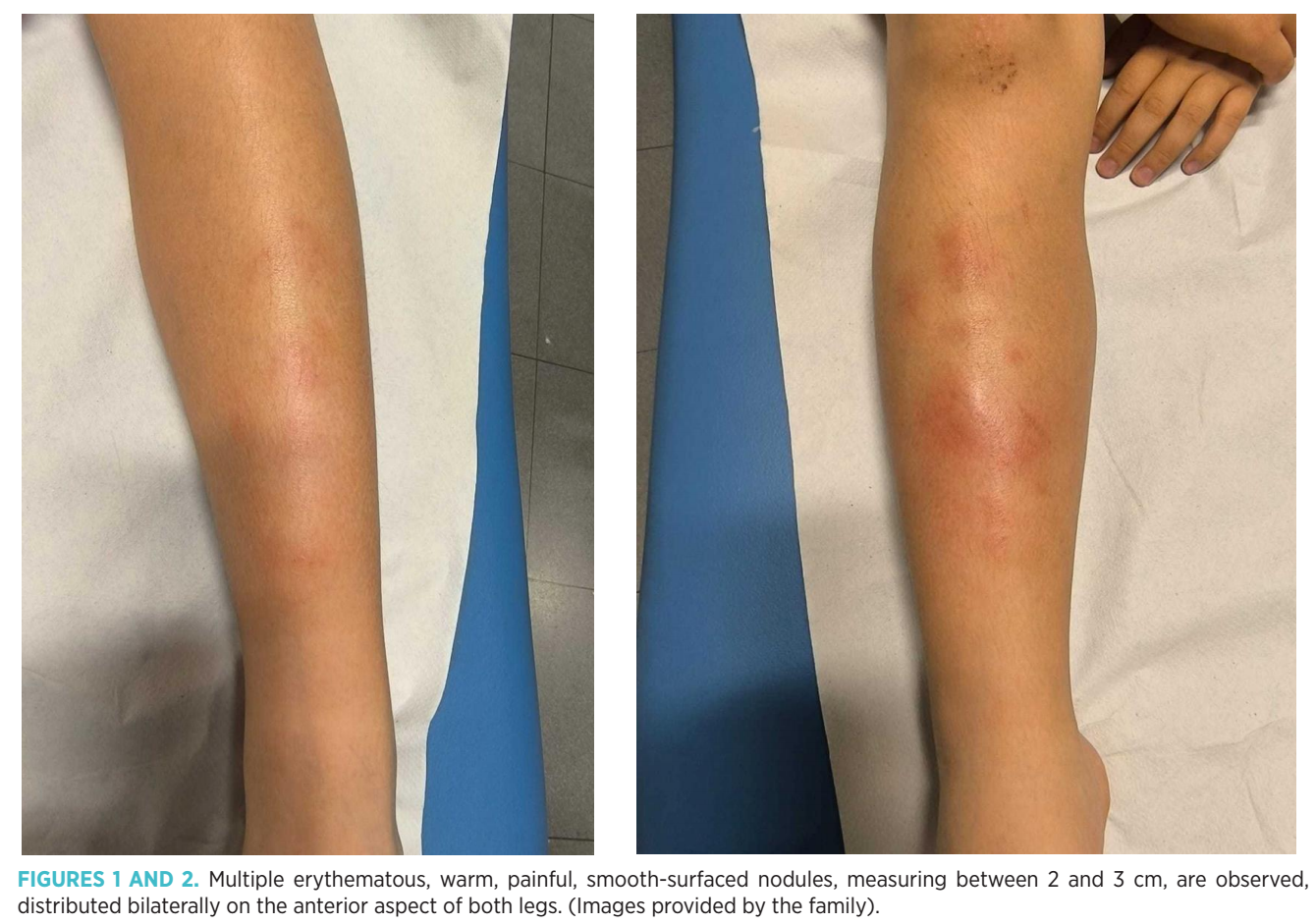
Given the suspicion of erythema nodosum, blood tests were performed, and complementary tests were requested for etiological study. The complete blood count, biochemistry (glucose, electrolytes, liver and renal profiles, and acute phase reactants), and urinalysis showed no alterations. The chest X-ray was normal. Pharyngeal culture for Streptococcus pyogenes and the Mantoux test were also negative.
Immunological tests showed a slight elevation of immunoglobulin A (serum IgA: 242 mg/dl), complement C3c (145 mg/dl) and C4 (44.7 mg/dl). ASLO and TSH determinations were within normal limits, and antinuclear antibodies (ANA) were negative.
Serologies detected positive IgM antibodies against the Epstein-Barr virus (VCA IgM). Serologies for Toxoplasma gondii, Cytomegalovirus, Borrelia burgdorferi, herpes simplex virus 1 and 2, Parvovirus B19, and Rickettsia conorii: negative.
The diagnosis of erythema nodosum was established in the Pediatric Emergency Department, requesting an urgent consultation with Dermatology to assess the need for a skin biopsy, which was deferred based on clinical evolution. Follow-up of non-urgent laboratory results, as well as cultures and serologies, was carried out in Pediatric outpatient clinics, also maintaining evolutionary control by Dermatology for the detection of possible complications.
The etiological study was carried out in a coordinated manner between the Pediatric Emergency Department, hospital Pediatrics, and the microbiology laboratory, which allowed identifying the cause of the condition without the need to resort to invasive procedures.
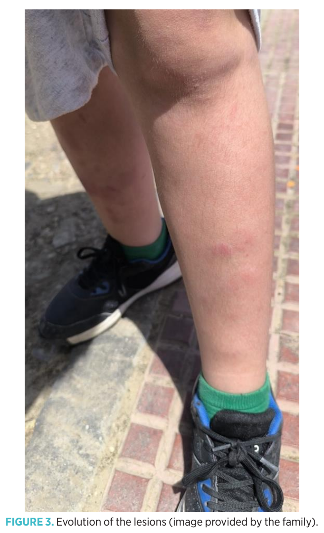
Symptomatic treatment with non-steroidal anti-inflammatory drugs and relative rest was indicated. The final clinical diagnosis was erythema nodosum associated with primary Epstein-Barr virus infection, with a favorable evolution and progressive resolution of the lesions in the subsequent weeks (Figure 3).
Discussion
Erythema nodosum is considered a septal panniculitis without vasculitis, generally mediated by a type IV delayed hypersensitivity immunological mechanism, which can be triggered by multiple infectious and non-infectious stimuli(6-7).
The diagnosis of erythema nodosum is fundamentally clinical and is based on the appearance of painful, erythematous, usually symmetrical subcutaneous nodules, located on the extensor surfaces of the lower limbs, in a patient with a good general condition(6-8). In the majority of pediatric cases, as in ours, skin biopsy is not necessary and is reserved for atypical presentations, ulcerated lesions, prolonged or recurrent evolution, or when there are diagnostic doubts with other panniculitis(6-7).
It is a rare entity in the pediatric population with no sex predilection(6). It is a benign, self-limiting process that usually resolves spontaneously in most cases within a period of 2 to 6 weeks, with an excellent long-term prognosis.
According to the literature, between 30 and 50% of cases are of idiopathic origin(6-7), with the causal agent not being identified, possibly due to the spontaneous resolution of initial symptoms and the absence of specific diagnostic markers(8).
The most prevalent known cause involved in EN during childhood is infectious, with Streptococcus pyogenes being the most frequent agent, followed by other viral infections, among which EBV infection stands out. However, in endemic areas, tuberculosis continues to be the most frequent infectious cause(3,9). There are other non-infectious causes, less common but relevant, such as sarcoidosis, rheumatological diseases, inflammatory bowel diseases, drug reactions, autoimmune disorders, pregnancy, and malignant neoplasms, which can also trigger this hypersensitivity reaction(3,11).
Zambrano-Mericq MJ and Lam JM published a review in 2023 that evidenced the association between EN and COVID-19, both after natural infection and after vaccination, including pediatric cases, which supports the need to consider this trigger in the etiological evaluation when no other apparent cause is identified(10).
In a study from Meyer Children’s Hospital in Florence (Italy), 68 pediatric patients diagnosed with EN were analyzed. A cause was identified in 55.9% of the cases, of which 42.6% were of infectious origin. The most frequent agents were EBV (22%), Streptococcus pyogenes (16.2%), Bartonella spp. (2.9%), Mycobacterium tuberculosis (1.5%) and Salmonella enteritidis (1.5%). Other causes included Crohn’s disease, celiac disease, Sjögren’s syndrome, and Hodgkin’s lymphoma. The mean age of the cohort was 8.8 years, with no significant sex differences. 25% presented comorbidities, mainly immunological or atopic. Approximately 26% had documented upper respiratory tract infection or gastroenteritis in the two weeks prior to the onset of symptoms, 10% had recent EBV infection, and 6% had a history of bacterial pharyngotonsillitis. A case with a history of meningococcus B vaccination one week before the onset of symptoms was also described(4) .
Although the association between EN and EBV is better documented in adults, it has also been described in the pediatric population. In the presented case, a primary EBV infection was confirmed as the triggering factor. The presence of previous symptoms, without evidence of bacterial infection or other clear etiology, guided the request for serological tests that allowed the etiological diagnosis to be established.
The management of erythema nodosum in the pediatric age group requires a multidisciplinary approach, where collaboration among emergency departments, pediatrics, dermatology, and microbiology is crucial to guide the etiological diagnosis and optimize the selection of complementary tests. Accurate identification of the causative agent allows for a more targeted management and avoids unnecessary treatments. In our case, this approach, supported by a detailed anamnesis and clinical judgment, allowed us to identify a primary Epstein-Barr virus infection as the trigger for the clinical picture.
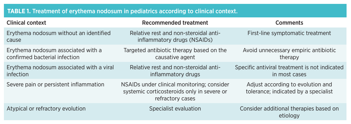
The treatment for erythema nodosum in the pediatric age group is primarily symptomatic. Relative rest and the use of nonsteroidal anti-inflammatory drugs are recommended, with ibuprofen being one of the most commonly used medications, within the usual anti-inflammatory pediatric range(6-8). The use of antibiotics is limited to those cases with documented bacterial infection(3-4,9). In exceptional cases of intense pain, prolonged evolution, or association with systemic disease, oral corticosteroids may be considered temporarily, evaluating individual risk-benefit(8) (Table 1).
Most cases of erythema nodosum in pediatrics resolve without sequelae, but in isolated situations, phenomena such as transient changes in the coloration of the lesions and persistence of pain for several days have been described. Exceptionally, encapsulated fat necrosis has been documented, a subcutaneous nodule of necrotic fat that can appear after inflammatory processes, including panniculitis such as erythema nodosum(12).
Outpatient clinical follow-up is recommended until complete resolution of the lesions. Reevaluation should be considered if symptoms persist, if there is a recurrence of episodes, or if systemic signs appear suggesting an underlying disease.
Comments
Identifying erythema nodosum as an inflammatory condition, rather than a localized infectious process, can reduce the unnecessary use of antibiotics and is crucial for ensuring appropriate management focused primarily on symptomatic relief. Although the etiological agent remains unidentified in a significant proportion of cases, when determined, it generally corresponds to an infectious agent, which highlights the importance of a complete diagnostic evaluation to guide treatment and follow-up.
Generally, this is a benign condition that tends to resolve spontaneously without long-term sequelae. This case underscores the importance of considering erythema nodosum in the differential diagnosis of the most common pediatric dermatitides(13), emphasizing the need for an exhaustive diagnostic approach to allow for accurate recognition and effective treatment.
Funding
No funding was received for the research, authorship, and/or publication of this article.
Authors’ contributions
Javier Pérez López: María Ángeles García Herrero: Writing - review and editing, Supervision
Carmen Moreno Solera: Writing - review and editing, Supervision
Elena Márquez Cáceres: Writing - review and editing, Supervision
Conflicts of interest
The authors declare that there is no commercial or financial conflict of interest for this research.
Ethical declarations
This work (clinical case, case series, etc.) includes patient data/images, and the authors confirm that they have obtained their express informed consent for its publication.
Use of artificial intelligence tools
The authors declare that no artificial intelligence tools (such as ChatGPT, Copilot, Gemini, or others) have been used in the writing, analysis, or review of this article.
References
- Requena L, Requena C. Eritema nodoso. Dermatol Online J. 2002;8(1):4.
- Agüero Orgaz D, Cubero Sobrados MC, Martín Gordo O. Eritema nodoso, a propósito de un caso. Rev Pediatr Aten Primaria. 2009;11:451–456.
- Urbaneja Rodríguez E, Garrote Molpeceres R. Eritema nodoso en pediatría. Protoc Diagn Ter Pediatr. 2020;2:285–293.
- Abu-Rumeileh S, Barbati F, Indolfi G, Trapani S. Erythema nodosum in children: a cohort study from a tertiary pediatric hospital in Italy. Eur J Pediatr. 2023 Apr;182(4):1803–1810. Available from: https://doi.org/10.1007/s00431-023-04872-x. Erratum in: Eur J Pediatr. 2024 May;183(5):2509. Disponible en: doi: 10.1007/s00431-024-05452-3.
- Alvarez Blanco O, Alvarez Montañana P, Mascuñán Díaz I. Eritema nudoso secundario a una infección enteral por Yersinia enterocolitica [Yersinia enterocolitica enteral infection-induced nodular erythema]. An Pediatr (Barc). 2008 Sep;69(3):282–283.
- Hafsi W, Badri T. Erythema nodosum. StatPearls [Internet]. Treasure Island (FL): StatPearls Publishing; 2025 Nov 28 [citado 2025 Aug 4]. Available from: https://www.ncbi.nlm.nih.gov/books/NBK556069/.
- Trapani S, Rubino C, Lodi L, Resti M, Indolfi G. Erythema nodosum in children: a narrative review and a practical approach. Children (Basel). 2022 Apr 4;9(4):511. Available from: https://doi.org/10.3390/children9040511.
- Pérez Garza DM, Chavez Alvarez S, Ocampo Candiani J, Gomez Flores M. Erythema nodosum: a practical approach and diagnostic algorithm. Am J Clin Dermatol. 2021 Mar;22(3):367–378. Available from: https://doi.org/10.1007/s40257-021-00607-7.
- Laborada J, Cohen PR. Tuberculosis-Associated Erythema Nodosum. Cureus. 2021 Dec 5;13(12):e20184. Available from: https://doi.org/10.7759/cureus.20184. PMID: 35004007; PMCID: PMC8723782.
- Zambrano-Mericq MJ, Lam JM. Erythema nodosum associated with COVID-19 infection: a pediatric case report and review of the literature. Pediatr Dermatol. 2023 Jan;40(1):166–170. Available from: https://doi.org/10.1111/pde.15096.
- Schwartz RA, Nervi SJ. Erythema nodosum: a sign of systemic disease. Am Fam Physician. 2007;75:695–700.
- Ahn SK, Lee BJ, Lee SH, Lee WS. Nodular cystic fat necrosis in a patient with erythema nodosum. Clin Exp Dermatol. 1995;20(3):263–265. Available from: https://doi.org/10.1111/j.1365-2230.1995.tb01319.x.
- Silva Rico JC, Torres Hinojal MC. Exantemas en la infancia. Pediatr Integral. 2014;XVIII(1):22–36.
Explorar
Artículos
Volúmenes
Sobre la revista
Más leídos
Emergencias Pediátricas
Emergencias Pediátricas
Emergencias Pediátricas
1º Congreso Mundial de Emergencias Pediátricas
Emergencias Pediátricas
Una nueva mirada a la vía aérea pediátrica: secuencia de intubación diferida como alternativa segura en situaciones de urgencias
Emergencias Pediátricas